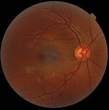
Fundus (eye)
The gaze is into the camera, so in each picture the macula is in the center of the image, and the optic disc is located towards the nose.
The major differences noted among the "higher" primate species [clarification needed] were size and regularity of the border of macular area, size and shape of the optic disc, apparent 'texturing' of retina, and pigmentation of retina.
Medical signs that can be detected from observation of eye fundus (generally by funduscopy) include hemorrhages, exudates, cotton wool spots, blood vessel abnormalities (tortuosity, pulsation and new vessels) and pigmentation.
[3] Arteriolar constriction, seen as "silver wiring", and vascular tortuosities are seen in hypertensive retinopathy.
The eye's fundus is the only part of the human body where the microcirculation can be observed directly.