In light microscopy, artifacts may be produced by air bubbles trapped under the slide's cover slip.
Different techniques including freeze-fracturing and cell fractionation may be used to overcome the problems of artifacts.
[1] A crush artifact is an artificial elongation and distortion seen in histopathology and cytopathology studies, presumably because of iatrogenic compression of tissues.
[4] In projectional radiography, visual artifacts that can constitute disease mimics include jewelry, clothes and skin folds.
[7] In Magnetic resonance imaging, artifacts can be classified as patient-related, signal processing-dependent or hardware (machine)-related.
A screenshot of a Microsoft
Windows XP
application displaying a visual artifact with repeated frames
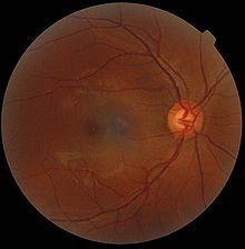
A
retinography
. The gray spot in the center is a shadow artifact.
Circular artifacts caused by backscatter from raindrops
Confocal laser scanning fluorescence micrograph of
thale cress
anther (part of
stamen
). The picture shows among other things a nice red flowing collar-like structure just below the anther. However, an intact thale cress stamen does not have such collar, this is a fixation artifact: the stamen has been cut below the picture frame, and
epidermis
(upper layer of cells) of stamen stalk has peeled off, forming a non-characteristic structure. Photo: Heiti Paves from
Tallinn University of Technology
.