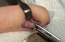
Glomus tumor
[5] Histologically, glomus tumors are made up of an afferent arteriole, anastomotic vessel, and collecting venule.
As stated above, these lesions should not be confused with paragangliomas, which were formerly also called glomus tumors in now-antiquated clinical usage.
Familial glomangiomas have been associated with a variety of deletions in the GLMN (glomulin) gene, and are inherited in an autosomal dominant manner, with incomplete penetrance.
In rare cases, the tumors may present in other body areas, such as the gastric antrum or glans penis.
There is one report of widespread metastases of a malignant glomus tumor involving the skin, lungs, jejunum, liver, spleen, and lymph nodes.
[10] The probable misdiagnosis of many of these lesions as hemangiomas or venous malformations also makes an accurate assessment of incidence difficult.
