Teratoma
A teratoma is a tumor made up of several types of tissue, such as hair, muscle, teeth, or bone.
[6][10] Teratomas occur in the coccyx in about one in 30,000 newborns, making them one of the most common tumors in this age group.
Immature teratoma has one of the lowest rates of somatic mutation of any tumor type and results from one of five mechanisms of meiotic failure.
[19] Gliomatosis peritoneii, which presents as a deposition of mature glial cells in the peritoneum, is almost exclusively seen in conjunction with cases of ovarian teratoma.
[19] A dermoid cyst is a mature cystic teratoma containing hair (sometimes very abundant) and other structures characteristic of normal skin and other tissues derived from the ectoderm.
Both forms may contain or appear to contain complete organ systems, even major body parts, such as a torso or limbs.
Fetus in fetu differs from fetiform teratoma in having an apparent spine and bilateral symmetry.
In many cases, the fetus in fetu is reported to occupy a fluid-filled cyst within a mature teratoma.
[25][26] Regardless of whether fetus in fetu and fetiform teratoma are one entity or two, they are distinct from and not to be confused with ectopic pregnancy.
A struma ovarii (also known as goitre of the ovary or ovarian goiter) is a rare form of mature teratoma that contains mostly thyroid tissue.
Teratomas of embryonal origin are most often found in babies at birth, in young children, and, since the advent of ultrasound imaging, in fetuses.
Because these teratomas project from the fetal body into the surrounding amniotic fluid, they can be seen during routine prenatal ultrasound exams.
[28][29] Teratomas are not dangerous for the fetus unless either a mass effect occurs or a large amount of blood flows through the tumor (known as vascular steal).
The mass effect frequently consists of obstruction of normal passage of fluids from surrounding organs.
Teratoma rarely include more complicated body parts such as teeth, brain matter,[30] eyes,[31][32] or torso.
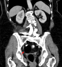
Ovarian teratomas often present with abdominal or pelvic pain, caused by torsion of the ovary or irritation of its ligaments.
Patients develop a multistage illness that progresses from psychosis, memory deficits, seizures, and language disintegration into a state of unresponsiveness with catatonic features often associated with abnormal movements, and autonomic and breathing instability.
(Maternal serum alpha-fetoprotein is a useful screening test for other fetal conditions, including Down syndrome, spina bifida, and abdominal wall defects such as gastroschisis.)
[39][40] Squamous cell carcinoma has been found in a mature cystic teratoma at the time of initial surgery.
[41] A grade 1 immature teratoma that appears to be benign (e.g., because AFP is not elevated) has a much higher risk of malignancy, and requires adequate follow-up.
Extraspinal ependymoma, usually considered to be a glioma (a type of nongerm cell tumor), may be an unusual form of mature teratoma.
Teratomas that are in surgically inaccessible locations, or are very complex, or are likely to be malignant (due to late discovery and/or treatment) sometimes are treated first with chemotherapy.
A UK study of 351 infants and children diagnosed with "benign" teratoma reported 227 with MT, 124 with IT.
Some teratomas secrete thyroxine, in some cases to such a degree that it can lead to clinical hyperthyroidism in the patient.
Adequate follow-up requires close observation, involving repeated physical examination, scanning (ultrasound, MRI, or CT), and measurement of AFP and/or βhCG.
[citation needed] Ovarian teratomas have been reported in mares,[57] mountain lions,[58][59] and canines.
[64] Because differentiated human pluripotent stem cells are being developed as the basis for numerous regenerative medicine therapies, there is concern that residual undifferentiated stem cells could lead to teratoma formation in injected patients, and researchers are working to develop methods to address this concern.