Dirofilaria immitis
[2] The definitive host is the dog, but it can also infect cats, wolves, coyotes, jackals, foxes, ferrets, bears, seals, sea lions and, under rare circumstances, humans.
[4] In cases involving advanced worm infestation, adult heartworms may migrate to the right heart and the pulmonary artery.
Although at one time confined to the southern United States, heartworm has now spread to nearly all locations where its mosquito vector is found.
[5] Transmission of the parasite occurs in all of the United States (cases have even been reported in Alaska), and the warmer regions of Canada.
The highest infection rates are found within 150 miles (240 km) of the coast from Texas to New Jersey, and along the Mississippi River and its major tributaries.
[4] It has also been found in South America,[6] southern Europe,[7][8] Southeast Asia,[9] the Middle East,[10] Australia, Korea, and Japan.
Between 75 and 120 days after infection, these immature heartworms then enter the bloodstream and are carried through the heart to reside in the pulmonary artery.
[15] By seven months after infection, the adult worms have mated and the females begin giving birth to live young, called microfilariae.
[16] The microfilariae circulate in the bloodstream for as long as two years, and are ingested by bloodsucking mosquitos, where development occurs and the cycle repeats.
Rarely, migrating heartworm larvae get "lost" and end up in aberrant sites, such as the eye, brain, or an artery in the leg, which results in unusual symptoms such as blindness, seizures, and lameness, but normally, until the larvae mature and congregate inside the heart, they produce no symptoms or signs of illness.
The inflammation occurring at the die-off of adult heartworms or larvae is in part due to the release of Wolbachia bacteria or protein into the tissues.
Treating heartworm-positive animals with an antibiotic such as doxycycline to remove Wolbachia may prove to be beneficial as it does for the filariae that cause elephantiasis,[22] but further studies are necessary.
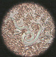
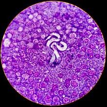
The Modified Knott's test is the best method of visual examination when determining presence of microfilaria because it preserves their morphology and size.
[4] Immunodiagnostics (ELISA, lateral flow immunoassay, rapid immunomigration techniques) to detect heartworm antigen in the host's blood are now regularly used.
Before the worms can be treated, however, the dog's heart, liver, and kidney function must be evaluated to determine the risks of treatment.
[30] It has a greater efficacy and fewer side effects than the previously used drug thiacetarsamide, sold as Caparsolate, which makes it a safer alternative for dogs with late-stage infections.
[citation needed] After treatment, the dog must rest, and exercise is to be heavily reduced for several weeks so as to give its body sufficient time to absorb the dead worms without ill effect.
Otherwise, if the dog is under exertion, dead worms may break loose and travel to the lungs, potentially causing respiratory failure and sudden death.
Once heartworm tests are negative and no surviving worm is detected, the treatment is considered a success, and the patient is effectively cured.
[citation needed] Surgical removal of the adult heartworms as a form of treatment may also be indicated, especially in advanced cases with substantial heart involvement and damage.
[citation needed] Preventive drugs are highly effective, and when regularly administered, have been shown to protect more than 99% of dogs and cats from heartworm.
If a mosquito feeds on a heartworm positive dog on a repellent, they do not live long enough for the microfilaria they ingested to molt into the infective L3 larva.
[19] Annual heartworm testing is highly recommended for pet owners who choose to use minimal dosing schedules.
A reaction has been identified in cats: heartworm-associated respiratory disease, which can occur three to four months after the initial infection, and is caused by the presence of the L5 larvae in the vessels.
[40] Obstruction of pulmonary arteries due to emboli from dying worms is more likely to be fatal in cats than dogs because of less collateral circulation and fewer vessels.
[16] Acute heartworm disease in cats can result in shock, vomiting, diarrhea, fainting, and sudden death.
Heartworm-associated respiratory disease can be found in cats that never develop adult heartworms and therefore never have a positive antigen test.
X-rays of the chest of a heartworm-infected cat may show an increased width of the pulmonary arteries and focal or diffuse opacities in the lungs.
[citation needed] Arsenic compounds have been used for heartworm adulticide treatment in cats, as well as dogs, but seem more likely to cause pulmonary reactions.
Several hundred cases of subcutaneous infections in humans have been reported in Europe, but these are almost always caused by another closely related parasite, Dirofilaria repens, rather than the dog heartworm.