Circulatory system
Many invertebrates such as arthropods have an open circulatory system with a heart that pumps a hemolymph which returns via the body cavity rather than via blood vessels.
An average adult contains five to six quarts (roughly 4.7 to 5.7 liters) of blood, accounting for approximately 7% of their total body weight.
[13] The heart pumps blood to all parts of the body providing nutrients and oxygen to every cell, and removing waste products.
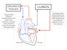
The left heart pumps oxygenated blood returned from the lungs to the rest of the body in the systemic circulation.
The blood that is returned to the right atrium is deoxygenated (poor in oxygen) and passed into the right ventricle to be pumped through the pulmonary artery to the lungs for re-oxygenation and removal of carbon dioxide.
The systemic circulation is a circuit loop that delivers oxygenated blood from the left heart to the rest of the body through the aorta.
[14] Oxygenated blood enters the systemic circulation when leaving the left ventricle, via the aortic semilunar valve.
The aorta arches and gives branches supplying the upper part of the body after passing through the aortic opening of the diaphragm at the level of thoracic ten vertebra, it enters the abdomen.
In humans, the only significant example is the hepatic portal vein which combines from capillaries around the gastrointestinal tract where the blood absorbs the various products of digestion; rather than leading directly back to the heart, the hepatic portal vein branches into a second capillary system in the liver.
The heart itself is supplied with oxygen and nutrients through a small "loop" of the systemic circulation and derives very little from the blood contained within the four chambers.
The neurovascular unit, composed of various cells and vasculature channels within the brain, regulates the flow of blood to activated neurons in order to satisfy their high energy demands.
[25] The human arterial system originates from the aortic arches and from the dorsal aortae starting from week 4 of embryonic life.
About 98.5% of the oxygen in a sample of arterial blood in a healthy human, breathing air at sea-level pressure, is chemically combined with hemoglobin molecules.
It is also a risk factor for acute coronary syndromes, which are diseases that are characterised by a sudden deficit of oxygenated blood to the heart tissue.
Cardiovascular diseases may also be congenital in nature, such as heart defects or persistent fetal circulation, where the circulatory changes that are supposed to happen after birth do not.
Hemolymph is composed of water, inorganic salts (mostly sodium, chloride, potassium, magnesium, and calcium), and organic compounds (mostly carbohydrates, proteins, and lipids).
[citation needed] In amphibians and most reptiles, a double circulatory system is used, but the heart is not always completely separated into two pumps.
[citation needed] In reptiles, the ventricular septum of the heart is incomplete and the pulmonary artery is equipped with a sphincter muscle.
[30] Double circulatory systems permit blood to be repressurized after returning from the lungs, speeding up delivery of oxygen to tissues.
Instead, a muscular pharynx leads to an extensively branched digestive system that facilitates direct diffusion of nutrients to all cells.
The flatworm's dorso-ventrally flattened body shape also restricts the distance of any cell from the digestive system or the exterior of the organism.
[citation needed] In the 6th century BCE, the knowledge of circulation of vital fluids through the body was known to the Ayurvedic physician Sushruta in ancient India.
[citation needed] In 1025, The Canon of Medicine by the Persian physician, Avicenna, "erroneously accepted the Greek notion regarding the existence of a hole in the ventricular septum by which the blood traveled between the ventricles."
Despite this, Avicenna "correctly wrote on the cardiac cycles and valvular function", and "had a vision of blood circulation" in his Treatise on Pulse.
The blood from the right chamber must flow through the vena arteriosa (pulmonary artery) to the lungs, spread through its substances, be mingled there with air, pass through the arteria venosa (pulmonary vein) to reach the left chamber of the heart and there form the vital spirit...In addition, Ibn al-Nafis had an insight into what later became a larger theory of the capillary circulation.
He stated that "there must be small communications or pores (manafidh in Arabic) between the pulmonary artery and vein," a prediction that preceded the discovery of the capillary system by more than 400 years.
Michael Servetus was the first European to describe the function of pulmonary circulation, although his achievement was not widely recognized at the time, for a few reasons.
Only three copies of the book survived but these remained hidden for decades, the rest were burned shortly after its publication in 1553 because of persecution of Servetus by religious authorities.
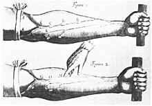
[citation needed] Finally, the English physician William Harvey, a pupil of Hieronymus Fabricius (who had earlier described the valves of the veins without recognizing their function), performed a sequence of experiments and published his Exercitatio Anatomica de Motu Cordis et Sanguinis in Animalibus in 1628, which "demonstrated that there had to be a direct connection between the venous and arterial systems throughout the body, and not just the lungs.
Most importantly, he argued that the beat of the heart produced a continuous circulation of blood through minute connections at the extremities of the body.