Human embryonic development
The genetic material of the sperm and egg then combine to form the single cell zygote and the germinal stage of development commences.
The germinal stage refers to the time from fertilization through the development of the early embryo until implantation is completed in the uterus.
The entire process of embryogenesis involves coordinated spatial and temporal changes in gene expression, cell growth, and cellular differentiation.
Fertilization takes place when the spermatozoon has successfully entered the ovum and the two sets of genetic material carried by the gametes fuse together, resulting in the zygote (a single diploid cell).
[5] Initially, the dividing cells, called blastomeres (blastos Greek for sprout), are undifferentiated and aggregated into a sphere enclosed within the zona pellucida of the ovum.
The hatching of the human embryo is supported by proteases secreted by the cells of the blastocyst, which digest proteins of the zona pellucida, giving rise to a hole.
The inner cell mass will give rise to the pre-embryo,[9] the amnion, yolk sac and allantois, while the fetal part of the placenta will form from the outer trophoblast layer.
The zona pellucida ultimately disappears completely, and the now exposed cells of the trophoblast allow the blastocyst to attach itself to the endometrium, where it will implant.
[11] The syncytiotrophoblast will grow and will enter a phase called lacunar stage, in which some vacuoles will appear and be filled by blood in the following days.
[12][13] Subsequently, new cells derived from yolk sac will be established between trophoblast and exocoelomic membrane and will give rise to extra-embryonic mesoderm, which will form the chorionic cavity.
This lining of the uterine cavity (or womb) is now known as the decidua, and it produces a great number of large decidual cells in its increased interglandular tissue.
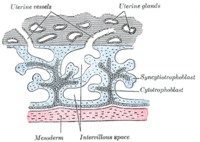
The syncytiotrophoblast implants the blastocyst in the decidual epithelium by projections of chorionic villi, forming the embryonic part of the placenta.
Progesterone enriches the uterus with a thick lining of blood vessels and capillaries so that it can oxygenate and sustain the developing embryo.
[11][15] Arteries in the decidua are remodelled to increase the maternal blood flow into the intervillous spaces of the placenta, allowing gas exchange and the transfer of nutrients to the embryo.
The primitive streak, a linear collection of cells formed by the migrating epiblast, appears, and this marks the beginning of gastrulation, which takes place around the seventeenth day (week 3) after fertilization.
The epiblast in that region moves down into the streak at the location of the primitive pit where the process called ingression, which leads to the formation of the mesoderm takes place.
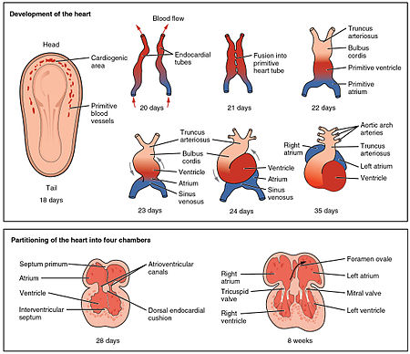
[17] The middle layer of mesoderm will give rise to the heart and the beginning of the circulatory system as well as the bones, muscles and kidneys.
The intermediate mesoderm gives rise to the urogenital tract and consists of cells that migrate from the middle region of the primitive line.
[11][15] The embryonic disc begins flat and round, but eventually elongates to have a wider cephalic part and narrow-shaped caudal end.
[10] At the beginning, the primitive line extends in cephalic direction and 18 days after fertilization returns caudally until it disappears.
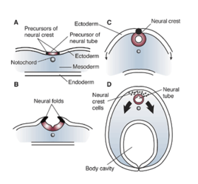
[11] Cranial and caudal neuropores become progressively smaller until they close completely (by day 26) forming the neural tube.
Blood islands develop outside the embryo, on the umbilical vesicle, allantois, connecting stalk, and chorion, from mesodermal hemangioblasts.
[21] Cardiac myoblasts and blood islands in the splanchnopleuric mesenchyme on each side of the neural plate give rise to the cardiogenic region.
Initially, all venous blood flows into the sinus venosus, and is propelled from tail to head to the truncus arteriosus.
The crescent shape prevents the complete closure of the atria allowing blood to be shunted from the right to the left atrium through the opening known as the ostium primum.
This closes with further development of the system but before it does, a second opening (the ostium secundum) begins to form in the upper atrium enabling the continued shunting of blood.
The septum primum is reduced to a small flap that acts as the valve of the foramen ovale and this remains until its closure at birth.
Between the fourth and seventh weeks of development, the urorectal septum divides the cloaca into the urogenital sinus and the anal canal.
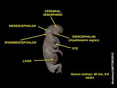
The mesenchyme that will form the dermis is derived from three sources: Late in the fourth week, the superior part of the neural tube bends ventrally as the cephalic flexure at the level of the future midbrain—the mesencephalon.
Six auricular hillocks, which are mesenchymal proliferations at the dorsal aspects of the first and second pharyngeal arches, form the auricle of the ear.