Human nose
[1] The nose is also made up of types of soft tissue such as skin, epithelia, mucous membrane, muscles, nerves, and blood vessels.
[3] The internal roof of the nasal cavity is composed of the horizontal, perforated cribriform plate of the ethmoid bone through which pass sensory fibres of the olfactory nerve.
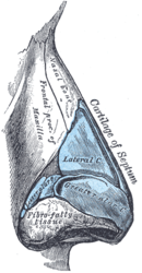
[3] At the peaks of the folds of the medial crura, they form the alar domes the tip-defining points of the nose, separated by a notch.
[13] There is a reinforcing structure known as the nasal scroll that resists internal collapse from airflow pressure generated by normal breathing.
[3] The anterior, and the posterior dilator naris, (the alar part of the nasalis muscle), give support to the nasal valves.
The lateral slip blends at the side of the upper lip with the levator labii superioris, and with the orbicularis oris.
The medial slip pulls the lateral crus upwards and modifies the curve of the furrow around the alae, and dilates the nostrils.
[21] At the back of the nasal cavity there are two openings, called choanae (also posterior nostrils), that give entrance to the nasopharynx, and rest of the respiratory tract.
[3] Excessive moisture as tears collected in the lacrimal sac travel down the nasolacrimal ducts where they drain into the inferior meatus in the nasal cavity.
The sudden change in the speed and pressure of the airflow creates turbulence that allows optimum contact with the respiratory epithelium for the necessary warming, moisturising, and filtering.
The levator labii superioris alaeque nasi is innervated by zygomatic and superior buccal branches of the facial nerve.
A plexiform network is formed in the lamina propria, by the bundles of axons that are surrounded by olfactory ensheathing cells.
In as many as twenty branches, the bundled axons cross the cribriform plate and enter the overlying olfactory bulb ending as glomeruli.
Postganglionic parasympathetic fibres derived from the pterygopalatine ganglion provide the secretomotor supply to the nasal mucous glands, and are distributed via branches of the maxillary nerves.
[3] In the early development of the embryo, neural crest cells migrate to form the mesenchymal tissue as ectomesenchyme of the pharyngeal arches.
The frontonasal process is a proliferation of mesenchyme in front of the brain vesicles,[48] and makes up the upper border of the stomadeum.
[49] During the seventh week the oronasal membrane ruptures and disintegrates to form an opening – the single primitive choana.
[51] Normal development is critical because the newborn infant breathes through the nose for the first six weeks, and any nasal blockage will need emergency treatment to clear.
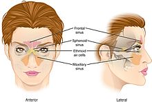
[52] The four pairs of paranasal sinuses – the maxillary, ethmoid, sphenoid, and frontal, develop from the nasal cavity as invaginations extending into their named bones.
Its main respiratory function is the supply and conditioning, by warming, moisturising and filtering of particulates of inhaled air.
[53] The internal structures and cavities, including the conchae and paranasal sinuses form an integrated system for the conditioning of the air breathed in through the nose.
[53] Sneezing is an important protective reflex action initiated by irritation of the nasal mucosa to expel unwanted particles through the mouth and nose.
[58] Variations in shape of the nose have been hypothesised to possibly be adaptive to regional differences in temperature and humidity, though they may also have been driven by other factors such as sexual selection.
This involves the lowering of the soft palate to produce nasal vowels and consonants by allowing air to escape from both the nose and the mouth.
[62] The large, hollow cavities of the paranasal sinuses act as resonating chambers that modify, and amplify speech and other vocal vibrations passing through them.
[83] Badly positioned alar cartilages lack proper support, and can affect the function of the external nasal valve.
[89] Similarly, "DIY nose lifts" in the form of re-usable cosmetic items have become popular and are sold in many Asian countries such as China, Japan, South Korea, Taiwan, Sri Lanka and Thailand.
The wiping of the nose with the hand, commonly referred to as the "allergic salute", is also mildly taboo and can result in the spreading of infections as well.
[100] Clive Finlayson of the Gibraltar Museum said the large Neanderthal noses were an adaptation to the cold,[101] Todd C. Rae of the American Museum of Natural History said primate and arctic animal studies have shown sinus size reduction in areas of extreme cold rather than enlargement in accordance with Allen's rule.
[102] Therefore, Todd C. Rae concludes that the design of the large and wide Neanderthal nose was evolved for the hotter climate of the Middle East and Africa and remained unchanged when they entered Europe.