Icerudivirus
The two strains were isolated from samples taken in 1994 from different solfataric fields in Iceland, the Kverkfjöll and Hveragerði, which are separated by a distance of 250 km.
[8] Virions are non-enveloped, consisting of a tube-like superhelix formed by dsDNA and the major structural protein, with plugs at each end to which three tail fibers are anchored.
These tail fibers appear to be involved in adsorption onto the host cell surface and are formed by one of the minor structural proteins.
[9] The structure revealed a previously unknown form of virion organization, in which the alpha-helical major capsid protein of SIRV2 wraps around the DNA, making it inaccessible to solvent.
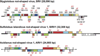
Although the sequences of the inverted terminal repeats of the rudiviruses are different, they all carry the motif AATTTAGGAATTTAGGAATTT near the genome ends, which may constitute a signal for the Holliday junction resolvase[10] and DNA replication.
[11] In contrast, at least 10% of its genes were predicted to have of different DNA binding motifs in the proteins they code and were assigned to be putative transcriptional regulators.
Virions are released from the host cell through a mechanism that involves the formation of specific cellular structures.