Iconic memory
[4] Classic experiments including Sperling's partial report paradigm as well as modern techniques continue to provide insight into the nature of this SM store.
[5] Natural observation of the light trail produced by glowing ember at the end of a quickly moving stick sparked the interest of researchers in the 1700s and 1800s.
In 1960, George Sperling began his classic partial-report experiments to confirm the existence of visual sensory memory and some of its characteristics including capacity and duration.
The second component is a longer-lasting memory store which represents a coded version of the visual image into post-categorical information.
The anterior superior temporal sulcus (STS), a part of the ventral stream, was found to be active in macaques during iconic memory tasks.
Iconic memory's role in change detection has been related to activation in the middle occipital gyrus (MOG).
MOG activation was found to persist for approximately 2000ms suggesting a possibility that iconic memory has a longer duration than what was currently thought.
[17] Iconic memory provides a smooth stream of visual information to the brain which can be extracted over an extended period of time by VSTM for consolidation into more stable forms.
One of iconic memory's key roles is involved with change detection of our visual environment which assists in the perception of motion.
The particular outcome depends on whether the two subsequent component images (i.e., the "icons") are meaningful only when isolated (masking) or only when superimposed (integration).
The brief representation in iconic memory is thought to play a key role in the ability to detect change in a visual scene.
The phenomenon of change blindness has provided insight into the nature of the iconic memory store and its role in vision.
[19] It has been suggested that iconic memory plays a role in providing continuity of experience during saccadic eye movements.
A similar phenomenon occurs during eye-blinks whereby both automatic and intentional blinking disrupts the information stored in iconic memory.
Iconic memory impairment in those with MCIs may be used as a predictor for the development of more severe deficits such as Alzheimer's disease and dementia later in life.
It reduces the active maintenance and storage of sensory information by altering transient neural responses during the initial stimulus processing stages.
[25] Elevated cortisol levels have also been associated with faster iconic memory decay and top-down processing impairment, putting individuals at a higher risk of developing Dementia and AD.
[26] Iconic memory formation has been previously described as attention-free and fleeting, however newer studies have shown that in fact it does require attention.
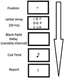
[2] In Sperling's initial experiments in 1960, observers were presented with a tachistoscopic visual stimulus for a brief period of time (50 ms) consisting of either a 3x3 or 3x4 array of alphanumeric characters such as: Recall was based on a cue which followed the offset of the stimulus and directed the subject to recall a specific line of letters from the initial display.
The whole report condition required participants to recall as many elements from the original display in their proper spatial locations as possible.
The partial report condition required participants to identify a subset of the characters from the visual display using cued recall.
Due to the fact that participants did not know which row would be cued for recall, performance in the partial report condition can be regarded as a random sample of an observer's memory for the entire display.
This type of sampling revealed that immediately after stimulus offset, participants could recall a given row (from a 3x3 grid of 9 letters) on 75% of trials, suggesting that 75% of the entire visual display (75% of 9-letters) was accessible to memory.
A small variation in Sperling's partial report procedure which yielded similar results was the use of a visual bar marker instead of an auditory tone as the retrieval cue.
Overall, experiments using partial report provided evidence for a rapidly decaying sensory trace lasting approximately 1000 ms after the offset of a display[2][30][31] The effects of masking were identified by the use of a circle presented around a letter as the cue for recall.