Immunocytochemistry
In cases where an immunopositive signal is found, ICC also allows researchers to determine which sub-cellular compartments are expressing the antigen.
This can be achieved by several methods: adherent cells may be grown on microscope slides, coverslips, or an optically suitable plastic support.
Reactions taking place in the nucleus can be more difficult, and the extracellular fluids can create unique obstacles in the performance of immunocytochemistry.
In this situation, permeabilizing cells using detergent (Triton X-100 or Tween-20) or choosing organic fixatives (acetone, methanol, or ethanol) becomes necessary.
There are many methods to obtain immunological detection on tissues, including those tied directly to primary antibodies or antisera.
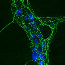
Alternatively the secondary antibody may be covalently linked to a fluorophore (FITC and Rhodamine are the most common) which is detected in a fluorescence or confocal microscope.
In this way immunofluorescence is a powerful technique when combined with confocal microscopy for studying the location of proteins and dynamic processes (exocytosis, endocytosis, etc.