Infarction
Infarction is tissue death (necrosis) due to inadequate blood supply to the affected area.
[1] The resulting lesion is referred to as an infarct[2][3] (from the Latin infarctus, "stuffed into").
[5] The blood vessel supplying the affected area of tissue may be blocked due to an obstruction in the vessel (e.g., an arterial embolus, thrombus, or atherosclerotic plaque), compressed by something outside of the vessel causing it to narrow (e.g., tumor, volvulus, or hernia), ruptured by trauma causing a loss of blood pressure downstream of the rupture, or vasoconstricted, which is the narrowing of the blood vessel by contraction of the muscle wall rather than an external force (e.g., cocaine vasoconstriction leading to myocardial infarction).
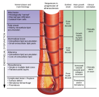
When the fibrous cap is degraded by metalloproteinases released from macrophages or by intravascular shear force from blood flow, subendothelial thrombogenic material (extracellular matrix) is exposed to circulating platelets and thrombus formation occurs on the vessel wall occluding blood flow.
Infarction in the brain requires first aid for stroke (using a protocol named F.A.S.T.