Inferior vena cava
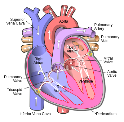
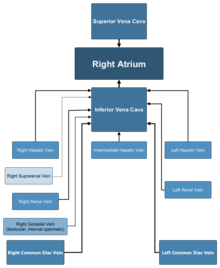
The inferior vena cava is a large vein that carries the deoxygenated blood from the lower and middle body into the right atrium of the heart.
Together, the venae cavae (in addition to the coronary sinus, which carries blood from the muscle of the heart itself) form the venous counterparts of the aorta.
It is a large retroperitoneal vein that lies posterior to the abdominal cavity and runs along the right side of the vertebral column.
The IVC is formed by the joining of the left and right common iliac veins and brings collected blood into the right atrium of the heart.
The inferior vena cava begins as the left and right common iliac veins behind the abdomen unite, at about the level of L5.
Ultrasound (US) systems are typically used to identify these variations; however, other techniques such as computed tomography (CT), which involves ionizing radiation, or magnetic resonance imaging (MRI), which is more costly, are often preferred due to the user-dependent nature of US analysis[5].
This may stem from the fact that it is easier to segment a closed cross-section than an open long-axis portion of the IVC, as the latter requires careful tracking of the region of interest[5].
Since the inferior vena cava is primarily a right-sided structure, unconscious pregnant women should be turned on to their left side (the recovery position), to relieve pressure on it and facilitate venous return[citation needed].
In rare cases, straining associated with defecation can lead to restricted blood flow through the IVC and result in syncope (fainting).