Intramembranous ossification
Intramembranous ossification is one of the two essential processes during fetal development of the gnathostome (excluding chondrichthyans such as sharks) skeletal system by which rudimentary bone tissue is created.
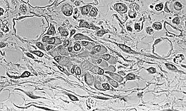
Before it begins to develop, the morphological characteristics of a MSC are: A small cell body with a few cell processes that are long and thin; a large, round nucleus with a prominent nucleolus that is surrounded by finely dispersed chromatin particles, giving the nucleus a clear appearance; and a small amount of Golgi apparatus, rough endoplasmic reticulum, mitochondria, and polyribosomes.
Furthermore, the mesenchymal stem cells are widely dispersed within an extracellular matrix that is devoid of every type of collagen, except for a few reticular fibrils.
[1] At this stage of development, changes in the morphology of the osteoprogenitor cells occur: Their shape becomes more columnar and the amount of Golgi apparatus and rough endoplasmic reticulum increases.
Much like spicules, the increasing growth of trabeculae result in interconnection and this network is called woven bone.
Embryologic mesenchymal cells (MSC) condense into layers of vascularized primitive connective tissue.
Osteogenic cells that originate from the periosteum increase appositional growth and a bone collar is formed.
When replacement to compact bone occurs, this blood vessel becomes the central canal of the osteon.