Iris (anatomy)

: irides or irises) is a thin, annular structure in the eye in most mammals and birds that is responsible for controlling the diameter and size of the pupil, and thus the amount of light reaching the retina.

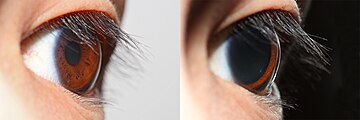
The high pigment content blocks light from passing through the iris to the retina, restricting it to the pupil.

The iris along with the anterior ciliary body provide a secondary pathway for aqueous humour to drain from the eye.

Many fish have neither, and, as a result, their irises are unable to dilate and contract, so that the pupil always remains of a fixed size.

The iris is usually strongly pigmented, with the color typically ranging between brown, hazel, green, gray, and blue.

The quantity of melanin pigment in the iris is one factor in determining the phenotypic eye color of an organism.

Most human irises also show a condensation of the brownish stromal melanin in the thin anterior border layer, which by its position has an overt influence on the overall color.

Abnormal clumping of melanosomes does occur in disease and may lead to irreversible changes in iris color (see heterochromia, below).

[citation needed] The optical mechanisms by which the nonpigmented stromal components influence eye color are complex, and many erroneous statements exist in the literature.

Interference is recognised by characteristic dependence of color on the angle of view, as seen in eyespots of some butterfly wings, although the chemical components remain the same.

White babies are usually born blue-eyed since no pigment is in the stroma, and their eyes appear blue due to scattering and selective absorption from the posterior epithelium.

Uncommon in humans, it is often an indicator of ocular disease, such as chronic iritis or diffuse iris melanoma, but may also occur as a normal variant.

Siberian Husky dogs show heterochromia,[12][better source needed] possibly analogous to the genetically determined Waardenburg syndrome of humans.

[13] Iridology (also known as iridodiagnosis) is an alternative medicine technique whose proponents believe that patterns, colors, and other characteristics of the iris can be examined to determine information about a patient's systemic health.

The iris (brown coloured portion of the eye) controls the size of the pupil by contracting the sphincter pupillae and dilator pupillae muscles.
Light micrograph of the iris near to the pupil. M. sph. sphincter muscle, L lens
Constriction of the pupil ( miosis ) observed by laser Doppler imaging reveals radial vessels of the iris.
A human eye demonstrating its owner's rare ability to voluntarily dilate and constrict his pupil on command, via voluntary control of his iris muscles.
Anterior chamber cross-section imaged by an SD-OCT .
Structure of the iris and surrounding parts showing the dilator and sphincter muscles ( dilator pupillae and sphincter pupillae ).
Human eye pigmentation in Europe
Among human phenotypes, blue-green-gray eyes are a relatively rare [ citation needed ] eye color and the exact color is often perceived to vary according to its surroundings.
A light brown iris with prominent limbal ring. Light brown irises contain pheomelanin.
Example of heterochromia – one eye of the subject is brown, the other hazel.
1:posterior segment 2:ora serrata 3:ciliary muscle 4:ciliary zonules 5:Schlemm's canal 6:pupil 7:anterior chamber 8:cornea 9:iris 10:lens cortex 11:lens nucleus 12:ciliary process 13:conjunctiva 14:inferior oblique muscule 15:inferior rectus muscule 16:medial rectus muscle 17:retinal arteries and veins 18:optic disc 19:dura mater 20:central retinal artery 21:central retinal vein 22:optic nerve 23:vorticose vein 24:bulbar sheath 25:macula 26:fovea 27:sclera 28:choroid 29:superior rectus muscle 30:retina 1: posterior segment 2: ora serrata 3: ciliary muscle 4: ciliary zonules 5: Schlemm's canal 6: pupil 7: anterior chamber 8: cornea 9: iris 10: lens cortex 11: lens nucleus 12: ciliary process 13: conjunctiva 14: inferior oblique muscule 15: inferior rectus muscule 16: medial rectus muscle 17: retinal arteries and veins 18: optic disc 19: dura mater 20: central retinal artery 21: central retinal vein 22: optic nerve 23: vorticose vein 24: bulbar sheath 25: macula 26: fovea 27: sclera 28: choroid 29: superior rectus muscle 30: retina
1:posterior segment 2:ora serrata 3:ciliary muscle 4:ciliary zonules 5:Schlemm's canal 6:pupil 7:anterior chamber 8:cornea 9:iris 10:lens cortex 11:lens nucleus 12:ciliary process 13:conjunctiva 14:inferior oblique muscule 15:inferior rectus muscule 16:medial rectus muscle 17:retinal arteries and veins 18:optic disc 19:dura mater 20:central retinal artery 21:central retinal vein 22:optic nerve 23:vorticose vein 24:bulbar sheath 25:macula 26:fovea 27:sclera 28:choroid 29:superior rectus muscle 30:retina