Laminotomy
[1] A laminotomy is less invasive than conventional vertebral column surgery techniques, such as laminectomy because it leaves more ligaments and muscles attached to the spinous process intact and it requires removing less bone from the vertebra.
[3] The vertebral arch is composed of several anatomical features in addition to laminae that must be taken into account when performing a laminotomy.
[3] The spinous process is located on the posterior or back side of the vertebra and serves as the attachment point for ligaments and muscles which support and stabilize the vertebral column.
[3] Each vertebra has two lateral bony projections called the transverse processes which are located on either side of the vertebral arch.
Transverse processes come into contact with the ribs and serve as attachment points for muscles and ligaments that stabilize the vertebral column.
[1] Both unilateral and bilateral laminotomies are performed in a shorter time period compared to a conventional laminectomy which takes over 100 minutes on average.
[1] The ligaments connecting the lamina of upper and lower vertebrae, known as Ligamenta flava are often removed or remodeled in this procedure to adjust for the small amount of bone lost.
[2] Using either a microscope or an endoscope to have a visual of the procedure, a small surgical drill is used to remove a part of bone from one or both laminae of the vertebrae.
The surgical tools are then navigated underneath the spinous process and across the spinal canal to reach the other lamina on the opposite side of the vertebra to perform a second laminotomy.
[2] Stenosis can be caused by old age or an injury to the vertebral column and usually requires a CT scan or MRI to diagnose.
[2] Performing a laminotomy can relieve pressure in the spinal canal caused by lumbar stenosis and therefore alleviate symptoms.
[2] Laminotomies are frequently used as a way to surgically repair a spinal disc herniation at any level of the vertebral column (cervical, thoracic, lumbar).
Some major complications that can occur are cerebrospinal fluid leaks, dural tears, infection, or epidural hematomas.
[5] A laminectomy is a more invasive method with the aim to decrease the total amount of pain and numbness associated with lumbar spinal stenosis.
Because a laminotomy does not damage the spinous process and critical ligaments, there is not as much muscle weakness, pain, and lumbar instability seen with laminectomies.
[4] Laminotomies are fairly new compared to laminectomies, and it involves using less invasive methods with precise instruments to minimize the risk of tissue damage.
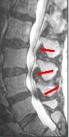
[5] For radiographic imaging, an x-ray is the least effective way to collect information when observing a patient with lumbar spinal stenosis.
The sharp contrast of the high power MRI outlines details in the vertebra that are critical when examining a patient with lumbar spinal stenosis who may need a laminotomy.
[2] A CT scan is not the most effective imaging technique when observing lumbar abnormalities, however it can supplement an MRI by detecting certain degenerative processes.
[2] Even though a CT scan can reveal these pertinent signs of lumbar spinal stenosis, it can sometimes give a cloudy image due to the shadowing of the tissue contrast.
[1] Other than static imaging processes, a CT scan can also be used for observing changes in spinal canal features before and after a laminotomy.
[citation needed] Minimally invasive procedures are a more common alternative due to the decreased risk of damaging significant muscle tissue.
Laminectomies have always been the gold standard when treating lumbar spinal stenosis, but recently, less invasive surgeries have emerged as a safer alternative treatment that helps maintain the postoperative structural integrity of the spine.
[4] A lumbar spinal fusion may be recommended when non-surgical treatment options for severe degenerative disc disease are ineffective.