Magnetoencephalography
[3] MEG signals were first measured by University of Illinois physicist David Cohen in 1968,[4] before the availability of the SQUID, using a copper induction coil as the detector.
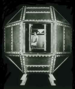
Later, Cohen built a much better shielded room at MIT, and used one of the first SQUID detectors, just developed by James E. Zimmerman, a researcher at Ford Motor Company,[5] to again measure MEG signals.
This was cumbersome, and, in the 1980s, MEG manufacturers began to arrange multiple sensors into arrays to cover a larger area of the head.
Recent developments attempt to increase portability of MEG scanners by using spin exchange relaxation-free (SERF) magnetometers.
The MEG (and EEG) signals derive from the net effect of ionic currents flowing in the dendrites of neurons during synaptic transmission.
[dubious – discuss] According to the right-hand rule, a current dipole gives rise to a magnetic field that points around the axis of its vector component.
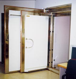
Appropriate magnetic shielding can be obtained by constructing rooms made of aluminium and mu-metal for reducing high-frequency and low-frequency noise, respectively.
This negatively feeds a DC amplifier through a low-pass network with a slow falloff to minimize positive feedback and oscillation.
The challenge posed by MEG is to determine the location of electric activity within the brain from the induced magnetic fields outside the head.
Localization algorithms make use of given source and head models to find a likely location for an underlying focal field generator.
If one's goal is to estimate the current density within the human brain with say a 5mm resolution then it is well established that the vast majority of the information needed to perform a unique inversion must come not from the magnetic field measurement but rather from the constraints applied to the problem.
A criticism of the use of this technique in clinical practice is that it produces colored areas with definite boundaries superimposed upon an MRI scan: the untrained viewer may not realize that the colors do not represent a physiological certainty, not because of the relatively low spatial resolution of MEG, but rather some inherent uncertainty in the probability cloud derived from statistical processes.
A widely accepted source-modeling technique for MEG involves calculating a set of equivalent current dipoles (ECDs), which assumes the underlying neuronal sources to be focal.
The matter is complicated by the fact that spatial resolution depends strongly on various parameters such as brain area, depth, orientation, number of sensors etc.
For creating functional maps of human cortex during more complex cognitive tasks, MEG is most often combined with fMRI, as the methods complement each other.
Neuronal (MEG) and hemodynamic fMRI data do not necessarily agree, in spite of the tight relationship between local field potentials (LFP) and blood oxygenation level-dependent (BOLD) signals.
There has been great success isolating unique responses in patients with schizophrenia, such as auditory gating deficits to human voices.
[20] Recent studies have reported successful classification of patients with multiple sclerosis, Alzheimer's disease, schizophrenia, Sjögren's syndrome, chronic alcoholism, facial pain and thalamocortical dysrhythmias.
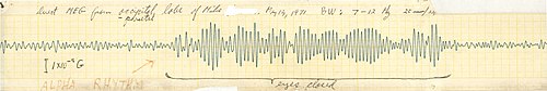
[23] Based on its perfect temporal resolution, magnetoencephalography (MEG) is now heavily used to study oscillatory activity in the brain, both in terms of local neural synchrony and cross-area synchronisation.
As an example for local neural synchrony, MEG has been used to investigate alpha rhythms in various targeted brain regions, such as in visual[24][25] or auditory cortex.
Direct cortical stimulation and somatosensory evoked potentials recorded on electrocorticography (ECoG) are considered the gold standard for localizing essential brain regions.
Noninvasive MEG localizations of the central sulcus obtained from somatosensory evoked magnetic fields show strong agreement with these invasive recordings.
Fetal recordings of cortical activity are feasible with a SARA device from a gestational age of approximately 25 weeks onward until birth.
[34] A third high density custom-made unit with similar whole abdomen coverage has been installed in 2002 at the University of Kansas Medical Center to assess fetal electrophysiology.
Such injuries are not easily diagnosed by other methods, as the symptoms (e.g. sleep disturbances, memory problems) overlap with those from frequent co-comorbidities such as post-traumatic stress disorder (PTSD).
Since the MEG signal is a direct measure of neuronal activity, its temporal resolution is comparable with that of intracranial electrodes.
MEG complements other brain activity measurement techniques such as electroencephalography (EEG), positron emission tomography (PET), and fMRI.
Finally, MEG is reference-free, while scalp EEG relies on a reference that, when active, makes interpretation of the data difficult.