Mechanical ventilation
Mechanical ventilation is termed invasive if it involves an instrument to create an airway that is placed inside the trachea.
There are many specific modes of mechanical ventilation, and their nomenclature has been revised over the decades as the technology has continually developed.The Greek physician Galen may have been the first to describe mechanical ventilation: "If you take a dead animal and blow air through its larynx [through a reed], you will fill its bronchi and watch its lungs attain the greatest distention."
In 1908, George Poe demonstrated his mechanical respirator by asphyxiating dogs and seemingly bringing them back to life.
Early ventilators were control style with no support breaths integrated into them and were limited to an inspiration to expiration ratio of 1:1.
It may, however, be used at home or in a nursing or rehabilitation institution for patients that have chronic illnesses that require long-term ventilatory assistance.
[12][13] Another well-documented complication is ventilator-associated lung injury which presents as acute respiratory distress syndrome.
One of the primary complications that presents in patients mechanically ventilated is acute lung injury (ALI)/acute respiratory distress syndrome (ARDS).
This is done by changing the mode to one where they have to trigger breaths and ventilatory support is only given to compensate for the added resistance of the endotracheal tube.
[27] A cuff leak test is done to detect if there is airway edema to show the chances of post-extubation stridor.
This phenomenon of respiration involves the physiologic concepts of air flow, tidal volume, compliance, resistance, and dead space.
Due to the anatomy of the human pharynx, larynx, and esophagus and the circumstances for which ventilation is needed, additional measures are required to secure the airway during positive-pressure ventilation in order to allow unimpeded passage of air into the trachea and avoid air passing into the esophagus and stomach.
It is not clear if clonidine is safe or effective to be used as a sedative for preterm and full term infants who require mechanical ventilation.
[32] In normal physiology, gas exchange of oxygen and carbon dioxide occurs at the level of the alveoli in the lungs.
If such complications are not present, other causes must be sought after, and positive end-expiratory pressure (PEEP) should be used to treat this intrapulmonary shunt.
[35] Further, this mode allows to use thin endotracheal tubes (~2 – 10 mm inner diameter) to ventilate a patient as expiration is actively supported.
[37] The design of the modern positive-pressure ventilators were based mainly on technical developments by the military during World War II to supply oxygen to fighter pilots in high altitude.
Positive pressure through manual supply of 50% oxygen through a tracheostomy tube led to a reduced mortality rate among patients with polio and respiratory paralysis.
However, because of the sheer amount of man-power required for such manual intervention, mechanical positive-pressure ventilators became increasingly popular.
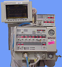
[2] Positive-pressure ventilators work by increasing the patient's airway pressure through an endotracheal or tracheostomy tube.
Then, the airway pressure drops to zero, and the elastic recoil of the chest wall and lungs push the tidal volume — the breath-out through passive exhalation.
[40] The prominent design of the smaller devices is known as the cuirass, a shell-like unit used to create negative pressure only to the chest using a combination of a fitting shell and a soft bladder.
In recent years this device has been manufactured using various-sized polycarbonate shells with multiple seals, and a high-pressure oscillation pump in order to carry out biphasic cuirass ventilation.
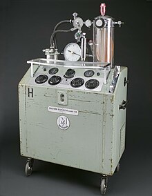
[2] The larger units have their origin in the iron lung, also known as the Drinker and Shaw tank, which was developed in 1928 by J.H Emerson Company and was one of the first negative-pressure machines used for long-term ventilation.
[3] The neck is sealed with a rubber gasket so that the patient's face (and airway) are exposed to the room air.
In the iron lung by means of a pump, the air is withdrawn mechanically to produce a vacuum inside the tank, thus creating negative pressure.
[3] Some of the problems with the full body design were such as being unable to control the inspiratory to expiratory ratio and the flow rate.
It works in conjunction with a separate CMV ventilator to add pulses of air to the control breaths and PEEP.
There are various procedures and mechanical devices that provide protection against airway collapse, air leakage, and aspiration: