Mitral regurgitation
[7] Individuals with chronic compensated MR may be asymptomatic for long periods of time, with a normal exercise tolerance and no evidence of heart failure.
Over time, however, there may be decompensation and patients can develop volume overload (congestive heart failure).
Symptoms of entry into a decompensated phase may include fatigue, shortness of breath particularly on exertion, and leg swelling.
The mitral component of the first heart sound is usually soft and with a laterally displaced apex beat,[6] often with heave.
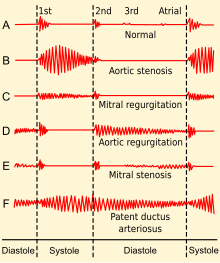
[7] The first heart sound is followed by a high-pitched holosystolic murmur at the apex, radiating to the back or clavicular area.
[8] Mitral regurgitation as a result of papillary muscle damage or rupture may be a complication of a heart attack and lead to cardiogenic shock.
[citation needed] The most common cause of MR in developed countries is mitral valve prolapse.
This can lead to the subsequent displacement of the papillary muscles and the dilatation of the mitral valve annulus.
[6] Mitral valve stenosis (MVS) can sometimes be a cause of mitral regurgitation (MR) in the sense that a stenotic valve (calcified and with restricted range of movement) allows backflow (regurgitation) if it is too stiff and misshapen to close completely.
Because the papillary muscles, chordae, and valve leaflets are usually normal in such conditions, it is also called functional mitral regurgitation.
In this phase, the left ventricle develops eccentric hypertrophy in order to better manage the larger than normal stroke volume.
A decreased ejection fraction in an individual with MR and no other cardiac abnormality should alert the physician that the disease may be in its decompensated phase.
The ECG may not show any of these findings in the setting of acute MR.[citation needed] The quantification of MR usually employs imaging studies such as echocardiography or magnetic resonance angiography of the heart.
[6] A transesophageal echocardiogram can give clearer images if needed as the back of the heart can also be viewed.
Methods that have been used to assess the regurgitant fraction in mitral regurgitation include echocardiography, cardiac catheterization, fast CT scan, and cardiac MRI.The echocardiographic technique to measure the regurgitant fraction is to determine the forward flow through the mitral valve (from the left atrium to the left ventricle) during ventricular diastole, and comparing it with the flow out of the left ventricle through the aortic valve in ventricular systole.
[citation needed] The treatment of MR depends on the acuteness of the disease and whether there are associated signs of hemodynamic compromise.
In general, medical therapy is non-curative and is used for mild-to-moderate regurgitation or in patients unable to tolerate surgery.
[15] In acute MR secondary to a mechanical defect in the heart (i.e., rupture of a papillary muscle or chordae tendineae), the treatment of choice is mitral valve surgery.
In the artificial chordae approach, ePTFE (expanded polytetrafluoroethylene, or Gore-Tex) sutures are used to replace the broken or stretched chordae tendonae, bringing the natural tissue back into the physiological position, thus restoring the natural anatomy of the valve.
With both techniques, an annuloplasty ring is typically secured to the annulus, or opening of the mitral valve, to provide additional structural support.
[23][24] Indications for surgery for chronic MR include signs of left ventricular dysfunction with ejection fraction less than 60%, severe pulmonary hypertension with pulmonary artery systolic pressure greater than 50 mmHg at rest or 60 mmHg during activity, and new-onset atrial fibrillation.
[citation needed] Significant mitral valve regurgitation has a prevalence of approximately 2% of the population, affecting males and females equally.