Nanoshell
[2] Nanoshells can be varied across a broad range of the light spectrum that spans the visible and near infrared regions.
Otherwise, a p-polarization is formed which gives a more strongly shifted plasmon energy causing a weaker interaction and coupling.
The production process described below was an experiment performed by Suhanya Duraiswamy and Saif A. Khan of the Department of Chemical and Biomolecular Engineering in Singapore.
The materials required for the production of the nanoshells are the following; Tetraethyl orthosilicate, ammonium hydroxide, hydroxylamine hydrochloride, 3-aminopropyl tris, hydrogentetrachloroaurate(III) trihydrate, tetrakis(hydroxymethyl) phosphonium chloride, sodium hydroxide, potassium carbonate, ethanol, Ultrapure water and glassware washed in aqua regia and rinsed thoroughly in water.
Microfluidic device patterns were fabricated onto silicon wafers by standard photolithography using negative photoresist SU-8 2050.
The microchannels were irreversibly bonded to a glass slide precoated with a thin layer of PDMS after a brief 35 s air plasma treatment.
The reason this method is revolutionary is that the size and relative thickness of the gold nanoshell can be controlled by changing the amount of time the reaction is allowed to take place as well as the concentration of the plating solution.
[7] The reason gold nanoparticles are used is due to their vivid optical properties which are controlled by their size, geometry, and their surface plasmons.
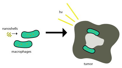
Under scattering, the gold-plated nano-particles become visible to imaging processes that are tuned to the correct wavelength which is dependent upon the size and geometry of the particles.
Under absorption, photothermal ablation occurs, which heats the nanoparticles and their immediate surroundings to temperatures capable of killing the cancer cells.
This is accomplished with minimal damage to cells in the body due to the utilization of the "water window" (the spectral range between 800 and 1300 nm).
This is accomplished because nanoparticle complexes are delivered inside of cells then undergo light induced release of DNA from their surface.