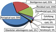
Odontogenic keratocyst
[6] Under The WHO/IARC classification, Odontogenic Keratocyst underwent the reclassification as it is no longer considered a neoplasm due to a lack of quality evidence regarding this hypothesis, especially with respect to clonality.
Within the Head and Neck pathology community there is still controversy surrounding the reclassification, with some pathologists still considering Odontogenic Keratocyst as a neoplasm in line with the previous classification.
However, bony expansion is uncommon as odontogenic keratocysts grow due to increased epithelial turnover rather than osmotic pressure.
[10] Sporadic (non-syndromic) and syndromic OKCs are associated with mutations in the gene PTCH found on chromosome 9q, which is part of the Hedgehog signaling pathway.
A third of OKCs show mutations in PTCH, resulting in the cyst epithelium undergoing highly proliferative activity.
Aspirational biopsy of odontogenic keratocysts contains a greasy fluid which is pale in colour and contains keratotic squames.
[2] Smaller and unilocular lesions resembling other types of cysts may require a biopsy to confirm the diagnosis.
[10] On a CT scan, the radiodensity of a keratocystic odontogenic tumour is about 30 Hounsfield units, which is about the same as ameloblastomas.
[14] Radiographs of odontogenic keratocysts show well-defined radiolucent areas with rounded or scalloped margins which are well demarcated.
Due to lack of expansion of the odontogenic keratocyst, the lesion can be very large when radiographically discovered.
Under the microscope, OKCs vaguely resemble keratinized squamous epithelium;[15] however, they lack rete ridges and often have an artifactual separation from their basement membrane.
Due to areas of focal inflammation, a larger biopsy is required for correct diagnosis of odontogenic keratocysts.
A substantial amount of odontogenic keratocysts also recur in the tooth-bearing area of the jaws, requiring attention from clinicians.
Due to high recurrence rate, late detection when the cyst has grown very large and causation by tumour suppressor gene inactivation, some have classified OKCs as benign neoplasms.