Oogenesis
In mammals, the first part of oogenesis starts in the germinal epithelium, which gives rise to the development of ovarian follicles, the functional unit of the ovary.
Mammalian oocytes are maintained in meiotic prophase arrest for a very long time—months in mice, years in humans.
Initially, the arrest is due to lack of sufficient cell cycle proteins to allow meiotic progression.
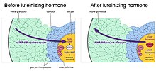
[5] Maintenance of meiotic arrest also depends on the presence of a multilayered complex of cells, known as a follicle, that surrounds the oocyte.
The granulosa cells produce a small molecule, cyclic GMP, that diffuses into the oocyte through the gap junctions.
[12] Two publications have challenged the belief that a finite number of oocytes are set around the time of birth generation in adult mammalian ovaries by putative germ cells in bone marrow and peripheral blood.
[13][14] The renewal of ovarian follicles from germline stem cells (originating from bone marrow and peripheral blood) has been reported in the postnatal mouse ovary.
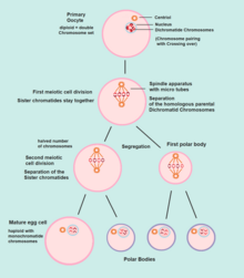
Meiosis I of ootidogenesis begins during embryonic development, but halts in the diplotene stage of prophase I until puberty.
If the egg is not fertilized, it is disintegrated and released (menstruation) and the secondary oocyte does not complete meiosis II (and does not become an ovum).
The function of forming polar bodies is to discard the extra haploid sets of chromosomes that have resulted as a consequence of meiosis.
[22] With this technique, cryopreserved ovarian tissue could possibly be used to make oocytes that can directly undergo in vitro fertilization.
[22] By definition it means, to recapitulate mammalian oogenesis and producing fertilizable oocytes in vitro.it is a complex process involving several different cell types, precise follicular cell-oocyte reciprocal interactions, a variety of nutrients and combinations of cytokines, and precise growth factors and hormones depending on the developmental stage.
[23] In 2016, two papers published by Morohaku et al. and Hikabe et al. reported in vitro procedures that appear to reproduce efficiently these conditions allowing for the production, completely in a dish, of a relatively large number of oocytes that are fertilizable and capable of giving rise to viable offspring in the mouse.
This technique can be mainly benefited in cancer patients where in today's condition their ovarian tissue is cryopreserved for preservation of fertility.
[25] However, homologous recombinational repair of DNA double-strand breaks mediated by BRCA1 and ATM weakens with age in oocytes of humans and other species.
Since older premenopausal women ordinarily have normal progeny, their capability for meiotic recombinational repair appears to be sufficient to prevent deterioration of their germline despite the reduction in ovarian reserve.
It has been suggested that such DNA damages may be removed, in large part, by mechanisms dependent on chromosome pairing, such as homologous recombination.
Oogenesis occurs within the embryo sac and leads to the formation of a single egg cell per ovule.


