Paracentesis
Anatomic landmarks, such as the midline linea alba approach, were traditionally used as reference points for needle insertion.
Phased array or curvilinear ultrasound transducers are typically used in the hospital and outpatient setting to identify ascites in the abdominal cavity.
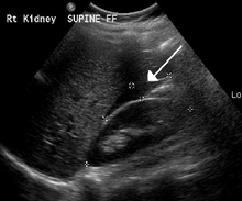
Morison's pouch (hepatorenal recess) is a common starting location in concordance with ultrasound FAST (focused assessment with sonography for trauma) exam.
Fluid collection can occur in a number of different locations and may be difficult to find, especially if the patient only exhibits a small volume of ascites.
Ultrasound guidance of the paracentesis can also be used as an additional safety measure to ensure the needle stays within the ascitic fluid and avoidance of important vessels within the abdominal cavity.
After cleaning the side of the abdomen with an antiseptic solution, the physician numbs a small area of skin and inserts a large-bore needle with a plastic sheath 2 to 5 cm (1 to 2 in) in length to reach the peritoneal (ascitic) fluid.
[7] There has been debate as to whether albumin administration confers benefit, but a recent 2016 meta-analysis concluded that it can reduce mortality after large-volume paracentesis significantly.
[8] However, for every end-point investigated, while albumin was favorable as compared to other agents (e.g., plasma expanders, vasoconstrictors), these were not statistically significant and the meta-analysis was limited by the quality of the studies—two of which that were in fact unsuitable—included in it.
A z-track is a technique that allows for decreased ascitic fluid leak following the paracentesis by displacing the needle tracks with respect to the epidermis and the peritoneum.