Pulmonary alveolus
[5] The acini are the basic units of respiration, with gas exchange taking place in all the alveoli present.
The respiratory bronchioles run for considerable lengths and become increasingly alveolated with side branches of alveolar ducts that become deeply lined with alveoli.
[5] A typical pair of human lungs contains about 480 million alveoli,[11] providing a total surface area for gas exchange of between 70 and 80 square metres.
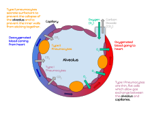
[12] An alveolus consists of an epithelial layer of simple squamous epithelium (very thin, flattened cells),[13] and an extracellular matrix surrounded by capillaries.
These cells are extremely thin – sometimes only 25 nm – the electron microscope was needed to prove that all alveoli are lined with epithelium.
This thin lining enables a fast diffusion of gas exchange between the air in the alveoli and the blood in the surrounding capillaries.
The cytoplasm in the thin portion contains pinocytotic vesicles which may play a role in the removal of small particulate contaminants from the outer surface.
Weaving between the capillaries and helping to support them is an extracellular matrix, a meshlike fabric of elastic and collagenous fibres.
This surfactant is a film of fatty substances, a group of phospholipids that reduce alveolar surface tension.
The fluid coating is produced by the body in order to facilitate the transfer of gases between blood and alveolar air, and the type II cells are typically found at the blood–air barrier.
[19][20] Type II cells start to develop at about 26 weeks of gestation, secreting small amounts of surfactant.
Alveolar macrophages also play a crucial role in immune responses against viral pathogens in the lungs.
[25] They secrete cytokines and chemokines, which recruit and activate other immune cells, initiate type I interferon signaling, and inhibit the nuclear export of viral genomes.
[25] Insufficient surfactant in the alveoli is one of the causes that can contribute to atelectasis (collapse of part or all of the lung).
[26] The severe condition of acute respiratory distress syndrome (ARDS) is caused by a deficiency or dysfunction of surfactant.
Cytokines and fluids are released into the alveolar cavity, interstitium, or both, in response to infection, causing the effective surface area of gas exchange to be reduced.
[35] Damaged capillaries from a contusion can cause blood and other fluids to accumulate in the tissue of the lung, impairing gas exchange.
An edema is usually caused by left ventricular heart failure, or by damage to the lung or its vasculature.