Respiratory tract
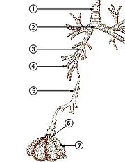
From the larynx, air moves into the trachea and down to the intersection known as the carina that branches to form the right and left primary (main) bronchi.
The diaphragm is also the main muscle of respiration involved in breathing, and is controlled by the sympathetic nervous system.
The lungs are encased in a serous membrane that folds in on itself to form the pleurae – a two-layered protective barrier.
From the bronchi, the dividing tubes become progressively smaller with an estimated 20 to 23 divisions before ending at an alveolus.
The tract consists of the nasal cavity and paranasal sinuses, the pharynx (nasopharynx, oropharynx and laryngopharynx) and sometimes includes the larynx.
Proximal divisions (those closest to the top of the tree, such as the bronchi) mainly function to transmit air to the lower airways.
Later divisions including the respiratory bronchiole, alveolar ducts, and alveoli, are specialized for gas exchange.
The trachea is the largest tube in the respiratory tract and consists of tracheal rings of hyaline cartilage.
This membrane secretes a small amount of fluid, allowing the lungs to move freely within the pleural cavity while expanding and contracting during breathing.
The alveoli are tiny air sacs in the lungs where gas exchange takes place.
[11] When the diaphragm contracts, a negative pressure is generated in the thorax and air rushes in to fill the cavity.
When the diaphragm relaxes, a positive pressure is generated in the thorax and air rushes out of the alveoli expelling the carbon dioxide.
There are glands and mucus produced by goblet cells in parts, as well as smooth muscle, elastin or cartilage.
Glands are abundant in the upper respiratory tract, but there are fewer lower down and they are absent starting at the bronchioles.
Respiration is the rhythmical process of breathing, in which air is drawn into the alveoli of the lungs via inhalation and subsequently expelled via exhalation.
Carbon dioxide (CO2) is transferred from returning blood back into gaseous form in the lungs and exhaled through the lower respiratory tract and then the upper, to complete the process of breathing.
As such, it has to be able to withstand suction pressures generated by the rhythmic expansion of the thoracic cavity that sucks air into the lungs.
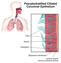
The epithelial lining of the upper respiratory tract is interspersed with goblet cells that secrete a protective mucus.
This helps to filter waste, which is eventually either swallowed into the highly acidic stomach environment or expelled via spitting.
The epithelium lining the respiratory tract is covered in small hairs called cilia.
Macrophages in the alveoli are part of the immune system which engulf and digest any inhaled harmful agents.
Hair in the nostrils plays a protective role, trapping particulate matter such as dust.
They also increase the surface area for particle deposition, improving the nose's ability to filter pathogens.
This causes difficulty in breathing and coughing as the lower respiratory tract tries to get rid of the fluid in the lungs.
Chronic bronchitis is common in smokers, because the tar from smoking accumulates over time, causing the lungs to work harder to repair themselves.
When a tobacco product is inhaled, the smoke paralyzes the cilia, causing mucus to enter the lungs.
Frequent smoking, over time, causes the cilia hairs to die and can no longer filter mucus.
The accumulation of this tar could eventually lead to lung cancer, or chronic obstructive pulmonary disease.
The decreased number of alveoli causes loss of oxygen supply to the lungs and an increased accumulation of carbon dioxide.