Adenocarcinoma of the lung
Once a lesion or tumor is identified with various imaging modalities, such as computed tomography (CT) or X-ray, a biopsy is required to confirm the diagnosis.
Surgical resection, chemotherapy, radiotherapy, targeted therapy and immunotherapy are used in attempt to eradicate the cancerous cells based upon these factors.
In nonsmokers, women and East Asians are more likely to present with symptoms of an underlying lung cancer at younger ages.
[10] Large scale studies such as The Cancer Genome Atlas (TCGA) have systematically characterized recurrent somatic alterations likely driving lung adenocarcinoma initiation and development.
[11] Since smoking is a strong mutagenic factor, lung adenocarcinoma is one of the tumor types with the highest number of mutations.
[2] Copy number amplifications in oncogenes such as TERT, Mdm2, EGFR, MET, and Myc have been reported, as well as deletions of tumor suppressor genes such as CDKN2A.
Adenocarcinoma of the lung develops in a step-wise progression as type II pneumocytes undergo consecutive molecular changes that disrupt normal cell regulation and turnover.
As more genetic mutations and dysregulation of normal cell signaling pathways accumulate, AAH can progress to adenocarcinoma in situ (AIS).
AIS lesions are classified as small tumors <3 cm with abnormal type II pneumocyte cell growth that is limited to the alveolar spaces i.e. without invasion into the stroma, pleura, or vasculature.
[15] Historically, there has been much debate in the most accurate method of describing adenocarcinoma of the lung and several revisions of classification systems have been published.
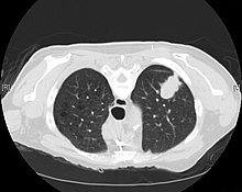
Computed tomography (CT) that is specifically aimed at evaluating lung cancer includes the chest and the upper abdomen.
This allows for evaluation of other relevant anatomic structures such as nearby lymph nodes, adrenal glands, liver, and bones which may show evidence of metastatic spread of disease.
It is also helpful for evaluating the extent of chest wall, diaphragmatic, brachial plexus (such as in the case of superior sulcus tumors), or spine involvement.
[4] If possible, a biopsy of any suspected lung tumor is performed to make a microscopic evaluation of the cells involved and is ultimately required to confirm diagnosis.
[30] The treatment of adenocarcinoma of the lung depends on several factors including stage, resectability, performance status, histology and genomic alterations acquired by the individual tumor.
Early stage (I, II and IIIA) lung adenocarcinomas are typically treated surgically to remove the tumor with pneumonectomy or lobectomy, if it is found to be resectable with imaging studies and biopsies and if the patient is considered able to tolerate surgery.
[34] For advanced (stage IV) and unresectable lung tumors, the first-line therapy is platinum-based doublet chemotherapy, combining cisplatin or carboplatin with another cytotoxic agent.
[3] Regimens strongly depend on each patient performance status and response, and when the risk of adverse events could worsen quality of life significantly, basic supportive care is more recommended.
Chemotherapy is also used as an adjuvant therapy following surgery to kill remaining cancer cells in patients with stage IIA, IIB and IIIA NSCLC.
[10] However, radiotherapy may be used as an adjuvant therapy for patients who have undergone a resection surgery to reduce the risk of lung cancer relapse.
It may also benefit inoperable tumors that are localized to the chest and be part of palliative care to improve quality of life in patients not responding to surgery or chemotherapy.
First-generation EGFR TKIs, including gefitinib and erlotinib, have been shown to be more effective in treating EGFR-mutated patients with respect to cytotoxic chemotherapy.
Most patients previously treated with crizotinib benefited from second-generation ALK inhibitors including ceritinib, alectinib and brigatinib.
[3][37] Howerver, the KEYNOTE-598 phase III trial has reported in 2021 that adding ipilimumab to pembrolizumab for NSCLC patients with PD-L1 tumor proportion score ≥50% does not confer any efficacy benefit, but may introduce greater toxicity.
Indeed, although smoking is still its strongest risk factor, lung adenocarcinoma is by far the most common among lifelong non-smokers (<100 cigarettes in a lifetime).