Root canal treatment
[1] Root canals, and their associated pulp chamber, are the physical hollows within a tooth that are naturally inhabited by nerve tissue, blood vessels and other cellular entities.
[9] There are several tests that can aid in the diagnosis of the dental pulp and the surrounding tissues: If a tooth is considered so threatened (because of decay, cracking, etc.)
The first, referred to as the standardized technique, was developed by Ingle in 1961, and had disadvantages such as the potential for loss of working length and inadvertent ledging, zipping or perforation.
These include the step-back, circumferential filing, incremental, anticurvature filing, step-down, double flare, crown-down-pressureless, balanced force, canal master, apical box, progressive enlargement, modified double flare, passive stepback, alternated rotary motions, and apical patency techniques.
[22] Certain irrigants, such as sodium hypochlorite and chlorhexidine, have proved to be effective antimicrobials in vitro[22] and are widely used during root canal therapy worldwide.
[24] The standard filling material is gutta-percha, a natural polymer prepared from latex from the percha tree (Palaquium gutta).
The standard endodontic technique involves inserting a gutta-percha cone (a "point") into the cleaned-out root canal along with a sealing cement.
[citation needed] Pain control can be difficult to achieve at times because of anesthetic inactivation by the acidity of the abscess around the tooth apex.
[citation needed] Some dentists may decide to temporarily fill the canal with calcium hydroxide paste in order to thoroughly sterilize the site.
This strong base is left in place for a week or more to disinfect and reduce inflammation in surrounding tissue, requiring the patient to return for a second or third visit to complete the procedure.
[26] Temporary filling-materials allow the creation of hermetic coronal-seals preventing from coronal microleakage (i.e. contamination of the root canal by bacteria); their presence over the entire time-period to fill the root canal and restore the tooth crown is mandatory, for increasing the probability of the endodontic-treatment success.
[27][28][29][30] However, these temporary filling-materials create coronal seals which only remain hermetic during less than 30 days in average (mainly because of the bacteria the saliva contains).
[citation needed] Molars and premolars that have had root canal therapy should be protected with a crown that covers the cusps of the tooth.
Anterior teeth typically do not require full coverage restorations after a root canal procedure, unless there is extensive tooth loss from decay or for esthetics or unusual occlusion.
If complex and expensive restorative dentistry is contemplated then ideally the contaminated gutta percha would be replaced in a retreatment procedure to minimise the risk of failure.
[39][40] Corticosteroid intra-oral injections were found to alleviate pain in the first 24 hours in patients with symptomatic irreversible pulp inflammation.
[41] Instruments may separate (break) during root canal treatment, meaning a portion of the metal file used during the procedure remains inside the tooth.
The occurrence of file separation depends on the narrowness, curvature, length, calcification and number of roots on the tooth being treated.
[42] The risk of endodontic files fracturing can be minimised by:[43] A sodium hypochlorite incident results in an immediate reaction of severe pain, followed by edema, haematoma and ecchymosis, as a consequence of the solution escaping the confines of the tooth and entering the periapical space.
[44] This may be caused iatrogenically by binding or excessive pressure on the irrigant syringe or it may occur if the tooth has an unusually large apical foramen.
[45] Tooth discoloration is common following root canal treatment; however, the exact causes for this are not completely understood.
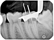
[citation needed] The X-ray in the right margin shows two adjacent teeth that had received bad root canal therapy.
[citation needed] Poor quality filling material or sealant may also cause root canal treatment to fail.
The perforation may be filled with a root repair material, such as one derived from natural cement called mineral trioxide aggregate (MTA).
[50] Endodontically treated teeth are prone to extraction mainly due to non-restorable carious destruction, other times due to the improper fit of the crown margins that encircles the tooth which lead to the ingress of bacteria,[51] and to a lesser extent to endodontic-related reasons such as endodontic failure, vertical root fracture, or perforation (procedural error).
People with special vulnerabilities, such as a recent prosthetic joint replacement, an unrepaired congenital heart defect, or immunocompromisation, may need to take antibiotics to protect from infection spreading during dental procedures.
A properly performed root canal treatment effectively removes the infected part of the pulp from the tooth.
Bacteremia (bacteria in the bloodstream) can be caused by many everyday activities, e.g. brushing teeth, but may also occur after any dental procedure which involves bleeding.
It is particularly likely after dental extractions due to the movement of the tooth and force needed to dislodge it, but endodontically treated teeth alone do not cause bacteremia or systemic disease.
[54] Endodontic therapy allows avoidance of disruption of the periodontal fiber, which helps with proprioception for occlusal feedback, a reflex important in preventing patients from chewing improperly and damaging the temporomandibular joint.