Rhabdomyolysis
Rhabdomyolysis (shortened as rhabdo) is a condition in which damaged skeletal muscle breaks down rapidly, often due to high intensity exercise over a short period.
[11] It is a significant problem for those injured in earthquakes, and relief efforts for such disasters often include medical teams equipped to treat survivors with rhabdomyolysis.
Milder forms may not cause any muscle symptoms, and the diagnosis is based on abnormal blood tests in the context of other problems.
[10] If the swelling is very rapid, as may happen with a crush injury after someone is released from under heavy collapsed debris, the movement of fluid from the bloodstream into damaged muscle may cause low blood pressure and shock.
[3][4][10] Release of the components of muscle tissue into the bloodstream causes electrolyte disturbances, which can lead to nausea, vomiting, confusion, coma or abnormal heart rate and rhythm.
[3][12] A second recognized complication is disseminated intravascular coagulation (DIC), a severe disruption in blood clotting that may lead to uncontrollable bleeding.
[16] Drugs of recreational use, including: alcohol, amphetamine, cocaine, heroin, ketamine and MDMA (ecstasy)[4][12] Recurrent or episodic rhabdomyolysis is commonly due to intrinsic muscle enzyme deficiencies, which are usually inherited and often appear during childhood.
[10] Neutrophil granulocytes—the most abundant type of white blood cell—enter the muscle tissue, producing an inflammatory reaction and releasing reactive oxygen species,[11] particularly after crush injury.
[18] As the kidneys reabsorb more water from the filtrate, myoglobin interacts with Tamm–Horsfall protein in the nephron to form casts (solid aggregates) that obstruct the normal flow of fluid; the condition is worsened further by high levels of uric acid and acidification of the filtrate, which increase cast formation.
[5] This enzyme is released by damaged muscle, and levels above 1000 U/L (5 times the upper limit of normal (ULN)) indicate rhabdomyolysis.
[5][11] CK concentrations rise steadily for 12 hours after the original muscle injury, remain elevated for 1–3 days and then fall gradually.
[19] Despite this, use of urine myoglobin measurement is not supported by evidence as it lacks specificity and the research studying its utility is of poor quality.
[4] Electrocardiography (ECG) may show whether the elevated potassium levels are affecting the conduction system of the heart, as suggested by the presence of T wave changes or broadening of the QRS complex.
[22][23] Disseminated intravascular coagulation, another complication of rhabdomyolysis and other forms of critical illness, may be suspected on the basis of unexpected bleeding or abnormalities in hematological tests, such as a decreasing platelet count or prolongation of the prothrombin time.
Initially this is done through the administration of generous amounts of intravenous fluids, usually isotonic saline (0.9% weight per volume sodium chloride solution).
[11][25] This will ensure sufficient circulating volume to deal with the muscle cell swelling (which typically commences when blood supply is restored), and to prevent the deposition of myoglobin in the kidneys.
[11][26] The rate of fluid administration may be altered to achieve a high urine output (200–300 mL/h in adults),[12][26] unless there are other reasons why this might lead to complications, such as a history of heart failure.
[26] While many sources recommend additional intravenous agents to reduce damage to the kidney, most of the evidence supporting this practice comes from animal studies, and is inconsistent and conflicting.
[10] The addition of bicarbonate to the intravenous fluids may alleviate acidosis (high acid level of the blood) and make the urine more alkaline to prevent cast formation in the kidneys;[10][12] evidence suggesting that bicarbonate has benefits above saline alone is limited, and it can worsen hypocalcemia by enhancing calcium and phosphate deposition in the tissues.
[26] Furosemide, a loop diuretic, is often used to ensure sufficient urine production,[4][11] but evidence that this prevents kidney failure is lacking.
[12] Temporary measures include the administration of calcium to protect against cardiac complications, insulin or salbutamol to redistribute potassium into cells, and infusions of bicarbonate solution.
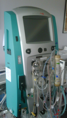
Hemodialysis, which is normally done several times a week in chronic kidney disease, is often required on a daily basis in rhabdomyolysis.
[4][10] The risk is higher in people with a history of illicit drug use, alcohol misuse or trauma when compared to muscle diseases, and it is particularly high if multiple contributing factors occur together.
The aftermath of the 1988 Spitak earthquake prompted the establishment, in 1995, of the Renal Disaster Relief Task Force, a working group of the International Society of Nephrology (a worldwide body of kidney experts).
Its volunteer doctors and nurses assisted for the first time in the 1999 İzmit earthquake in Turkey, where 17,480 people died, 5392 were hospitalized and 477 received dialysis, with positive results.
Treatment units are generally established outside the immediate disaster area, as aftershocks could potentially injure or kill staff and make equipment unusable.
[4] In modern times, early reports from the 1908 Messina earthquake and World War I on kidney failure after injury were followed by studies by London physicians Eric Bywaters and Desmond Beall, working at the Royal Postgraduate Medical School and the National Institute for Medical Research, on four victims of the Blitz in 1941.
[11][33] Already during the war, teams of doctors traveled to bombed areas to provide medical support, chiefly with intravenous fluids, as dialysis was not yet available.
[33] The prognosis of acute kidney failure improved markedly when dialysis was added to supportive treatment, which first happened during the 1950–1953 Korean War.
[36] Rhabdomyolysis affecting horses may also occur in outbreaks; these have been reported in many European countries, and later in Canada, Australia, and the United States.