Rectal prolapse
[5] It describes ulceration of the rectal lining caused by repeated frictional damage as the internal intussusception is forced into the anal canal during straining.
This classification also takes into account sphincter relaxation:[19] Rectal internal mucosal prolapse has been graded according to the level of descent of the intussusceptum, which was predictive of symptom severity:[20] The most widely used classification of internal rectal prolapse is according to the height on the rectal/sigmoid wall from which they originate and by whether the intussusceptum remains within the rectum or extends into the anal canal.
[3][5] Continent prolapse patients with slow transit constipation, and who are fit for surgery may benefit from subtotal colectomy with rectopexy.
[10] It may be used to assess for pelvic floor dyssenergia,[5] (anismus is a contraindication for certain surgeries, e.g. STARR), and these patients may benefit from post-operative biofeedback therapy.
[10] It may be used to evaluate incontinence, but there is disagreement about what relevance the results may show, as rarely do they mandate a change of surgical plan.
[21] When males are affected, they tend to be young and report significant bowel function symptoms, especially obstructed defecation,[5] or have a predisposing disorder (e.g., congenital anal atresia).
[5] Signs and symptoms include: Initially, the mass may protrude through the anal canal only during defecation and straining, and spontaneously return afterwards.
Shortly after the invention of defecography, In 1968 Broden and Snellman used cinedefecography to show that rectal prolapse begins as a circumferential intussusception of the rectum,[3][10] which slowly increases over time.
[21] This proved an older theory from the 18th century by John Hunter and Albrecht von Haller that this condition is essentially a full-thickness rectal intussusception, beginning about 3 inches above the dentate line and protruding externally.
[9] This excessive straining may be due to predisposing pelvic floor dysfunction (e.g. obstructed defecation) and anatomical factors:[10][21] Some authors question whether these abnormalities are the cause, or secondary to the prolapse.
[5] The assumed mechanism of obstructed defecation is by disruption to the rectum and anal canal's ability to contract and fully evacuate rectal contents.
The intussusceptum itself may mechanically obstruct the rectoanal lumen, creating a blockage that straining, anismus and colonic dysmotility exacerbate.
[30] The factors that result in a patient progressing from internal intussusception to a full thickness rectal prolapse remain unknown.
[5] Defecography studies demonstrated that degrees of internal intussusception are present in 40% of asymptomatic subjects, raising the possibility that it represents a normal variant in some, and may predispose patients to develop symptoms, or exacerbate other problems.
The perineal approach generally results in less post-operative pain and complications, and a reduced length of hospital stay.
[10] Alternatively, perineal procedures may be selected to reduce risk of nerve damage, for example in young male patients for whom sexual dysfunction may be a major concern.
Redundant rectal and sigmoid wall is removed and the new edge of colon is reconnected (anastomosed) with the anal canal with stitches or staples.
[3] Delorme Procedure This is a modification of the perineal rectosigmoidectomy, differing in that only the mucosa and submucosa are excised from the prolapsed segment, rather than full thickness resection.
"Mucosal proctectomy" was first discussed by Delorme in 1900,[10] now it is becoming more popular again as it has low morbidity and avoids an abdominal incision, while effectively repairing the prolapse.
Complications, including infection, urinary retention, bleeding, anastomotic dehiscence (opening of the stitched edges inside), rectal stricture (narrowing of the gut lumen), diarrhea, and fecal impaction occur in 6–32% of cases.
Complications include breakage of the encirclement material, fecal impaction, sepsis, and erosion into the skin or anal canal.
Symptom severity increases with the size of the prolapse, and whether it spontaneously reduces after defecation, requires manual reduction by the patient, or becomes irreducible.
Solitary rectal ulcer syndrome (SRUS, SRU), is a disorder of the rectum and anal canal, caused by straining and increased pressure during defecation.
[46] Pathological specimens of sections of rectal wall taken from SRUS patients show thickening and replacement of muscle with fibrous tissue and excess collagen.
Cystica profunda is characterized by formation of mucin cysts in the muscle layers of the gut lining, and it can occur anywhere along the gastrointestinal tract.
[18] Complications are uncommon, but include massive rectal bleeding, ulceration into the prostate gland or formation of a stricture.
[18][45] Some recommend biopsy as essential for diagnosis since ulcerations may not always be present, and others state defecography as the investigation of choice to diagnose SRUS.
Stopping straining during bowel movements, by use of correct posture, dietary fiber intake (possibly included bulk forming laxatives such as psyllium), stool softeners (e.g. polyethylene glycol,[63][64] and biofeedback retraining to coordinate pelvic floor during defecation.
Michelle Lhooq, writing for VICE, argues that rosebudding is an example of producers making 'extreme' content due to the easy availability of free pornography on the internet.
[70] Repeated rectal prolapses can cause bowel problems and anal leakage and therefore risk the health of pornographic actors or actresses who participate in them.

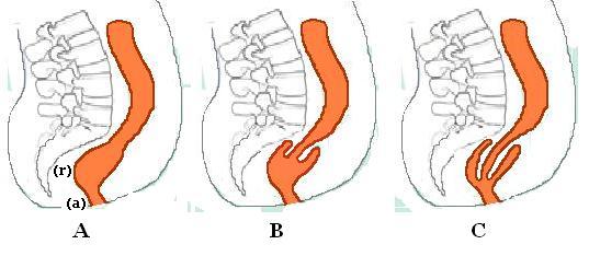
B. Recto-rectal intussusception
C. Recto-anal intussusception

