Single-photon emission computed tomography
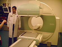
Single-photon emission computed tomography (SPECT, or less commonly, SPET) is a nuclear medicine tomographic imaging technique using gamma rays.
[1] It is very similar to conventional nuclear medicine planar imaging using a gamma camera (that is, scintigraphy),[2] but is able to provide true 3D information.
This information is typically presented as cross-sectional slices through the patient, but can be freely reformatted or manipulated as required.
The technique needs delivery of a gamma-emitting radioisotope (a radionuclide) into the patient, normally through injection into the bloodstream.
Usually, though, a marker radioisotope is attached to a specific ligand to create a radioligand, whose properties bind it to certain types of tissues.
Instead of just "taking a picture of anatomical structures", a SPECT scan monitors level of biological activity at each place in the 3-D region analyzed.
A computer is then used to apply a tomographic reconstruction algorithm to the multiple projections, yielding a 3-D data set.
This data set may then be manipulated to show thin slices along any chosen axis of the body, similar to those obtained from other tomographic techniques, such as magnetic resonance imaging (MRI), X-ray computed tomography (X-ray CT), and positron emission tomography (PET).
SPECT is similar to PET in its use of radioactive tracer material and detection of gamma rays.
Triggered by electrocardiogram (EKG) to obtain differential information about the heart in various parts of its cycle, gated myocardial SPECT can be used to obtain quantitative information about myocardial perfusion, thickness, and contractility of the myocardium during various parts of the cardiac cycle, and also to allow calculation of left ventricular ejection fraction, stroke volume, and cardiac output.
A cardiac specific radiopharmaceutical is administered, e.g., 99mTc-tetrofosmin (Myoview, GE healthcare), 99mTc-sestamibi (Cardiolite, Bristol-Myers Squibb) or Thallium-201 chloride.
SPECT imaging performed after stress reveals the distribution of the radiopharmaceutical, and therefore the relative blood flow to the different regions of the myocardium.
[5] This latter ability relates to SPECT's imaging of local metabolism of the brain, in which the patchy loss of cortical metabolism seen in multiple strokes differs clearly from the more even or "smooth" loss of non-occipital cortical brain function typical of Alzheimer's disease.
While 99mTc is extracted from relatively simple technetium-99m generators, which are delivered to hospitals and scanning centers weekly to supply fresh radioisotope, FDG PET relies on FDG, which is made in an expensive medical cyclotron and "hot-lab" (automated chemistry lab for radiopharmaceutical manufacture), and then delivered immediately to scanning sites because of the natural short 110-minute half-life of Fluorine-18.
Iterative reconstruction is an alternative algorithm that is growing in importance, as it is less sensitive to artifacts and can also correct for attenuation and depth dependent blurring.