Schistosoma mansoni
[1] Unlike other flukes (trematodes) in which sexes are not separate (monoecious), schistosomes are unique in that adults are divided into males and females, thus, gonochoric.
The posterior two-thirds of the body contains the vitelline glands and their winding canal, which unites with the oviduct a little before it reaches the ootype.
S. mansoni and other schistosomes are the only flukes or flatworms that exhibit sex separation as they exist as male and female individuals as in dioecious animals.
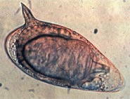
[22][23] The miracidium (from the Greek word μειράκιον, meirakion, meaning youth) is pear-shaped, and gradually elongates as it ages.
Epidermal plate is absent only at the extreme anterior called apical papilla, or terebratorium, which contains numerous sensory organelles.
[30] While hemoglobin is digested intracellularly, initiated by salivary gland enzymes, iron waste products cannot be used by the worms, and are typically discarded via regurgitation.
[31] Kasschau et al. (1995) tested the effect of temperature and pH on the ability of developing S. mansoni to lyse red blood cells.
[32] Ressurreicao et al. (2015) tested the roles of various protein kinases in the ability of the parasite to navigate its medium and locate a penetrable host surface.
[39] The cercaria emerge from the snail during daylight and they propel themselves in water with the aid of their bifurcated tail, actively seeking out their final host.
The paired worms move against the flow of blood to their final niche in the mesenteric circulation, where they begin egg production (>32 days).
The S. mansoni parasites are found predominantly in the small inferior mesenteric blood vessels surrounding the large intestine and caecal region of the host.
S. mansoni genome has increased protease families and deficiencies in lipid anabolism; which are attributed to its parasitic adaptation.
[51] In 2019, Ittiprasert, Brindley and colleagues employed programmed CRISPR/Cas9 knockout of the gene encoding the T2 ribonuclease of the egg of Schistosoma mansoni, advancing functional genomics and reverse genetics in the study of schistosomes, and platyhelminths generally
The hepatic form of the disease is the most important, granulomas here giving rise to fibrosis of the liver and hepatosplenomegaly in severe cases.
In the latter stages of the disease, the pathology is associated with collagen deposition and fibrosis, resulting in organ damage that may be only partially reversible.
[53] The granulomas formed around the eggs impair blood flow in the liver and, as a consequence, induce portal hypertension.
With time, collateral circulation is formed and the eggs disseminate into the lungs, where they cause more granulomas, pulmonary arteritis and, later, cor pulmonale.
This fibrosis occurs only many years after the infection and is presumed to be caused in part by soluble egg antigens and various immune cells that react to them.
Synthetic IL-13Rα2 given to mice has resulted in significant decreases in granuloma size, implicating IL-13Rα2 as an important target in schistosomiasis.
One important factor was the development of large reservoir of infection due to extensive schistosomiasis control programs that used intravenously administered tartar emetic since the 1960s.
The worms have many tools that help in this evasion, including the tegument, antioxidant proteins, and defenses against host membrane attack complex (MAC).
Targeting of this pathway with different inhibitors of the central antioxidant enzyme thioredoxin glutathione reductase (TGR) results in reduced viability of worms.
[59] Decay accelerating factor (DAF) protein is present on the parasite tegument and protects host cells by blocking formation of MAC.
[63] However, this is not frequently used in countries where the disease is common due to the cost of the equipment and the technical expertise required to run them.
Young children living in these areas are at greatest risk because of their tendency to swim and bathe in cercaria-infected waters longer than adults .
The intermediate hosts Biomphalaria snails are estimated to originate in South America 95–110 million years ago.
[72][73] A German physician Theodor Maximillian Bilharz was the first to discover the parasite in 1851, while working at Kasr el-Aini Hospital, a medical school in Cairo.
Manson identified lateral-spined eggs in the faeces of a colonial officer earlier posted to the West Indies, and concluded that there were two species of Schistosoma.
In 1908, a Brazilian physician Manuel Augusto Pirajá da Silva gave a complete description of male and female worms, including the lateral-spined eggs.
[82] The species identity was confirmed in 1907 by British parasitologist Robert Thomson Leiper,[79] identifying the specific snail host, and distinguishing the egg structure, thereby establishing the life cycle.