Scrotal ultrasound
Many of the disease processes, such as testicular torsion, epididymo-orchitis, and intratesticular tumor, produce the common symptom of pain at presentation, and differentiation of these conditions and disorders is important for determining the appropriate treatment.
High-resolution ultrasound aids in improved characterization of some intrascrotal lesions and suggests more specific diagnoses, resulting in more appropriate treatments and the avoidance of unnecessary operation.
A transverse image including all or a portion of both testicles in the field of view is obtained to allow side-to-side comparison of their sizes, echogenicity, and vascularity.
The testicle is surrounded by a dense white fibrous capsule, the tunica albuginea, which is often not visualized in the absence of intrascrotal fluid.
The appendix testis is a Müllerian duct remnant and consists of fibrous tissue and blood vessels within an envelope of columnar epithelium.
Seminomas occur in a slightly older age group when compared with other nonseminomatous tumor, with a peak incidence in the fourth and fifth decades.
Lymphatic spread to retroperitoneal lymph nodes and hematogenous metastases to lung, brain, or both are evident in about 25% of patients at the time of presentation.
Choriocarcinomas are composed of both cytotrophoblasts and syncytiotrophoblasts, with the latter responsible for the clinical elevation of human chorionic gonadotrophic hormone levels.
Many choriocarcinomas show extensive hemorrhagic necrosis in the central portion of the tumor; this appears as mixed cystic and solid components at ultrasound.
8], then the patient's age at presentation, symptoms, and medical history, as well as multiplicity and bilaterality of the lesions, are all important factors in making the appropriate diagnosis.
However, due to the presence of blood-testis barrier, chemotherapeutic agents are unable to reach the testis, hence in boys with acute lymphoblastic leukemia, testicular involvement is reported in 5% to 10% of patients, with the majority found during clinical remission.
Clinically, epidermoid cyst cannot be differentiated from other testicular tumors, typically presenting as a non-tender, palpable, solitary intratesticular mass.
[citation needed] However, these patterns, except the latter one, may be considered as non-specific as heterogeneous echotexture and shadowing calcification can also be detected in malignant testicular tumors.
Absence of vascular flow is another important feature that is helpful in differentiation of epidermoid cyst from other solid intratesticular lesions.
Hydrocele, either simple or complex is present and may be associated with:[citation needed] Leiomyomas are benign neoplasms that may arise from any structure or organ containing smooth muscle.
The majority of genitourinary leiomyomas are found in the renal capsule, but this tumor has also been reported in the epididymis, spermatic cord, and tunica albuginea.
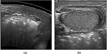
It can be divided into three types depending upon the site of origination and spread:[citation needed] At ultrasound, lipoma is a well–defined, homogeneous, hyperechoic paratesticular lesion of varying size [Fig.
Although the exact cause of this tumor is not completely understood, it is generally believed that these lesions represent a benign reactive proliferation of inflammatory and fibrous tissue, in response to chronic irritation.
[citation needed] At ultrasound, the findings of acute epididymitis include an enlarged hypoechoic or hyperechoic (presumably secondary to hemorrhage) epididymis [Fig.
[citation needed] Fournier gangrene is a polymicrobial necrotizing fasciitis involving the perineal, perianal, or genital regions and constitutes a true surgical emergency with a potentially high mortality rate.
[citation needed] The sonographic hallmark of Fournier gangrene is presence of subcutaneous gas within the thickened scrotal wall.
[citation needed] Histologically, testicular microlithiasis refers to the scattered laminated calcium deposits in the lumina of the seminiferous tubules.
[citation needed] The US appearance of varicocele consists of multiple, hypoechoic, serpiginous, tubular like structures of varying sizes larger than 2 mm in diameter that is usually best visualized superior or lateral to the testis [Fig.
[citation needed] Normally the testes begin its descent through the inguinal canal to the scrotum at 36 weeks’ of gestation and completed at birth.
[citation needed] Besides infertility, undescended testes carry an increased risk of malignancy even for the normally located contralateral testis.
Physical examination showed a small, firm nodule is palpable on the superior aspect of the testis and a bluish discoloration known as ‘‘blue dot’’ sign may be seen on the overlying skin.
The sonographic features of testicular appendiceal torsion include a circular mass with variable echogenicity located adjacent to the testis or epididymis [Fig.
30], reactive hydrocele and skin thickening of the scrotum is common, increased peripheral vascular flow may be found around the testicular appendage on color Doppler ultrasound.
A striated pattern of the testicle, radiating from its mediastinum, does not have clinical importance unless there are alarming symptoms or abnormal signal on Doppler ultrasonography.
[8] Ultrasound remains as the mainstay in scrotal imaging not only because of its high accuracy, excellent depiction of scrotal anatomy, low cost and wide availability, it is also useful in determining whether a mass is intra- or extra-testicular, thus providing us useful and valuable information to decide whether a mass is benign or malignant even though malignancy does occur in extratesticular tumors and vice versa.