Subarachnoid hemorrhage
[1] Symptoms may include a severe headache of rapid onset, vomiting, decreased level of consciousness, fever, weakness, numbness, and sometimes seizures.
[13] Oculomotor nerve abnormalities (affected eye looking downward and outward and inability to lift the eyelid on the same side) or palsy (loss of movement) may indicate bleeding from the posterior communicating artery.
SAH is a frequent occurrence in traumatic brain injury and carries a poor prognosis if it is associated with deterioration in the level of consciousness.
In these cases prognosis is poorer; however, it is unclear if this is a direct result of the SAH or whether the presence of subarachnoid blood is simply an indicator of a more severe head injury.
[4] Cocaine abuse and sickle cell anemia (usually in children) and, rarely, anticoagulant therapy, problems with blood clotting and pituitary apoplexy can also result in SAH.
Oxyhaemoglobin in cerebrospinal fluid (CSF) causes vasoconstriction by increasing free radicals, endothelin-1, prostaglandin and reducing the level of nitric oxide and prostacyclin.
[27] As only 10 percent of people admitted to the emergency department with a thunderclap headache are having an SAH, other possible causes are usually considered simultaneously, such as meningitis, migraine, and cerebral venous sinus thrombosis.
[30] The initial steps for evaluating a person with a suspected subarachnoid hemorrhage are obtaining a medical history and performing a physical examination.
The diagnosis cannot be made on clinical grounds alone and in general medical imaging and possibly a lumbar puncture is required to confirm or exclude bleeding.
[33] Lumbar puncture, in which cerebrospinal fluid (CSF) is removed from the subarachnoid space of the spinal canal using a hypodermic needle, shows evidence of bleeding in three percent of people in whom a non-contrast CT was found normal.
[4] A lumbar puncture or CT scan with contrast is therefore regarded as mandatory in people with suspected SAH when imaging is delayed to after six hours from the onset of symptoms and is negative.
[47] The World Federation of Neurosurgeons (WFNS) classification uses Glasgow coma score and focal neurological deficit to gauge severity of symptoms.
[52] On the basis of the ISUIA and other studies, it is now recommended that people are considered for preventive treatment only if they have a reasonable life expectancy and have aneurysms that are highly likely to rupture.
In general, pain control is restricted to less-sedating agents such as codeine, as sedation may impact on the mental status and thus interfere with the ability to monitor the level of consciousness.
[11] People with poor clinical grade on admission, acute neurologic deterioration, or progressive enlargement of ventricles on CT scan are, in general, indications for the placement of an external ventricular drain by a neurosurgeon.
[1] People whose CT scan shows a large hematoma, depressed level of consciousness, or focal neurologic signs may benefit from urgent surgical removal of the blood or occlusion of the bleeding site.
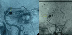
[23] The calcium channel blocker nimodipine when taken by mouth improves outcome if given between the fourth and twenty-first day after the bleeding, even if it does not reduce the amount of vasospasm detected on angiography.
[63] Nimodipine is readily authorized in the form of tablets and solution for infusion for the prevention and treatment of complications due to vasospasm following subarachnoid hemorrhage.
If the level of consciousness is decreased, drainage of the excess fluid is performed by therapeutic lumbar puncture, extraventricular drain (a temporary device inserted into one of the ventricles), or occasionally a permanent shunt.
[11] Fluctuations in blood pressure and electrolyte imbalance, as well as pneumonia and cardiac decompensation occur in about half the hospitalized persons with SAH and may worsen prognosis.
[81] Factors that carry a worse prognosis during the hospital stay include occurrence of delayed ischemia resulting from vasospasm, development of intracerebral hematoma, or intraventricular hemorrhage (bleeding into the ventricles of the brain) and presence of fever on the eighth day of admission.
[22] Perimesencephalic SAH (bleeding around the mesencephalon in the brain), however, has a very low rate of rebleeding or delayed ischemia, and the prognosis of this subtype is excellent.
[23] It is difficult to isolate the effects of SAH from those of other aspects of traumatic brain injury; it is unknown whether the presence of subarachnoid blood actually worsens the prognosis or whether it is merely a sign that a significant trauma has occurred.
For example, having two copies of ApoE4 (a variant of the gene encoding apolipoprotein E that also plays a role in Alzheimer's disease) seems to increase risk for delayed ischemia and a worse outcome.
[88] Aneurysmal subarachnoid hemorrhage may lead to damage of the hypothalamus and the pituitary gland, two areas of the brain that play a central role in hormonal regulation and production.
[90] Risk of SAH is about 25 percent higher in women over 55 compared to men the same age, probably reflecting the hormonal changes that result from the menopause, such as a decrease in estrogen levels.
[90] Genetics may play a role in a person's disposition to SAH; risk is increased three- to fivefold in first-degree relatives of people having had a subarachnoid hemorrhage.
[84] There is likely an inverse relationship between total serum cholesterol and the risk of non-traumatic SAH, though confirmation of this association is hindered by a lack of studies.
[52] While the clinical picture of subarachnoid hemorrhage may have been recognized by Hippocrates, the existence of cerebral aneurysms and the fact that they could rupture was not established until the 18th century.
[96] The 1980s saw the introduction of triple H therapy[76] as a treatment for delayed ischemia due to vasospasm, and trials with nimodipine[69][97] in an attempt to prevent this complication.