Synovial chondromatosis
Once it reaches transitional the loose bodies become apparent with X-ray in greater than 70% of cases, with MRI often showing where xray fails.
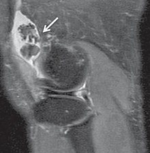
[2] Rare and little known, with currently no known cure, the disease gradually forms blisters in the thin flexible membrane of the synovium, which calcify and enlarge.
The affected tissue will show up as a semi-solid mass in an MRI scan, final diagnosis is usually confirmed by taking a biopsy.
However, online communities for synovial chondromatosis patients have yielded a stark contrast, with equal representation from both genders and members diagnosed as young as late teenage/early 20s.
Secondary synovial chondromatosis is the more common form and often occurs when there is pre-existent osteoarthritis, rheumatoid arthritis, osteonecrosis, osteochondritis dissecans, neuropathic osteoarthropathy (which often occurs in diabetic individuals), tuberculosis, or osteochondral fractures (torn cartilage covering the end of a bone in a joint) in the affected individual.
Synovectomies are normally carried out by shaving the lining of the knee but there are other ways of achieving this by either freezing the synovium or by the use of radiation treatment.