Taenia saginata
As hermaphrodites, each body segment called proglottid has complete sets of both male and female reproductive systems.
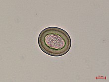
From humans, embryonated eggs, called oncospheres, are released with faeces and are transmitted to cattle through contaminated fodder.
The inside of each mature proglottid is filled with muscular layers and complete male and female reproductive systems, including the tubular unbranched uterus, ovary, genital pore, testes, and vitelline gland.
Oncospheres enter the duodenum, the anterior portion of the small intestine, and hatch there under the influence of gastric juices.
The larvae can move to all parts of the body by the general circulatory system, and finally settle in skeletal muscles within 70 days.
Inside the tissue, they cast off their hooks and instead develop a protective cuticular shell, called the cyst.
The inner membrane of the cysticercus soon develops numerous protoscolices (small scolices) that are invertedly attached to the inner surface.
Once reaching the jejunum, the inverted scolex becomes evaginated to the exterior under stimuli from the digestive enzymes of the host.
In each mature proglottid, self-fertilisation produces zygotes, which divide and differentiate into embryonated eggs called oncospheres.
These oncospheres in an external environment can remain viable for several days to weeks in sewage, rivers, and pastures.
[3][6][7] The disease is relatively common in Africa, some parts of Eastern Europe, the Philippines, and Latin America.
[8] This parasite is found anywhere where beef is eaten, including countries such as the United States, with strict federal sanitation policies.
[11] Taenia saginata has been reported as a cause of gallbladder perforation if left untreated in some cases.
[13] The Taenia saginata remains asymptomatic due to the fact the organism does not present cysticerci in humans.
Since it is difficult to diagnose using eggs alone, looking at the scolex or the gravid proglottids can help identify it as Taenia saginata.
[6] Proglottids sometimes trickle down the thighs of infected humans and are visible with unaided eye, so can aid with identification.
[2] Differentiation of the species of Taenia, such as T. solium and T. asiatica, is notoriously difficult because of their close morphological resemblance, and their eggs are more or less identical.
Identification often requires histological observation of the uterine branches and PCR detection of ribosomal 5.8S gene.