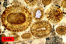
Thin section
A thin sliver of rock is cut from the sample with a diamond saw and ground optically flat.
It is then mounted on a glass slide and then ground smooth using progressively finer abrasive grit until the sample is only 30 μm thick.
When placed between two polarizing filters set at right angles to each other, the optical properties of the minerals in the thin section alter the colour and intensity of the light as seen by the viewer.
Thin sections are prepared in order to investigate the optical properties of the minerals in the rock.
In thin section, when viewed in plane polarized light (PPL), quartz is colorless with low relief and no cleavage.
This technique has been used to study the microstructure of fine-grained carbonates such as the Lochseitenkalk mylonite in which the matrix grains are less than 5 μm in size.