Regeneration (biology)
Regeneration in biology is the process of renewal, restoration, and tissue growth that makes genomes, cells, organisms, and ecosystems resilient to natural fluctuations or events that cause disturbance or damage.
[7][8] Regeneration in biology, however, mainly refers to the morphogenic processes that characterize the phenotypic plasticity of traits allowing multi-cellular organisms to repair and maintain the integrity of their physiological and morphological states.
[10] The cells underneath this cap then begin to rapidly divide and form a cone shaped end to the amputation known as a blastema.
[9] A planarian parent, for example, will constrict, split in the middle, and each half generates a new end to form two clones of the original.
The case of autotomy, for example, serves as a defensive function as the animal detaches a limb or tail to avoid capture.
Following a disturbance, such as a fire or pest outbreak in a forest, pioneering species will occupy, compete for space, and establish themselves in the newly opened habitat.
[20][21] Pattern formation in the morphogenesis of an animal is regulated by genetic induction factors that put cells to work after damage has occurred.
Neural cells, for example, express growth-associated proteins, such as GAP-43, tubulin, actin, an array of novel neuropeptides, and cytokines that induce a cellular physiological response to regenerate from the damage.
Later, this conserved signaling pathway was also found to be essential for regeneration of many mammalian tissues, including heart, liver, skin, and lung, and intestine.
[36] For example, Chaetopterus variopedatus and Branchiomma nigromaculata can regenerate both anterior and posterior body parts after latitudinal bisection.
[37] The relationship between somatic and germline stem cell regeneration has been studied at the molecular level in the annelid Capitella teleta.
[44] Alejandro Sanchez-Alvarado and Philip Newmark transformed planarians into a model genetic organism in the beginning of the 20th century to study the molecular mechanisms underlying regeneration in these animals.
[55] Epidermal cells continue to migrate over the WE, resulting in a thickened, specialized signaling center called the apical epithelial cap (AEC).
[56] Over the next several days there are changes in the underlying stump tissues that result in the formation of a blastema (a mass of dedifferentiated proliferating cells).
[57] Motor neurons, muscle, and blood vessels grow with the regenerated limb, and reestablish the connections that were present prior to amputation.
Researchers at Australian Regenerative Medicine Institute at Monash University have published that when macrophages, which eat up material debris,[59] were removed, salamanders lost their ability to regenerate and formed scarred tissue instead.
[61] In spite of the historically few researchers studying limb regeneration, remarkable progress has been made recently in establishing the neotenous amphibian the axolotl (Ambystoma mexicanum) as a model genetic organism.
This progress has been facilitated by advances in genomics, bioinformatics, and somatic cell transgenesis in other fields, that have created the opportunity to investigate the mechanisms of important biological properties, such as limb regeneration, in the axolotl.
Located at the University of Kentucky, the AGSC is dedicated to supplying genetically well-characterized axolotl embryos, larvae, and adults to laboratories throughout the United States and abroad.
[67] Hydra is a genus of freshwater polyp in the phylum Cnidaria with highly proliferative stem cells that gives them the ability to regenerate their entire body.
In the enhancer regions that are activated during head regeneration, a set of transcription factor motifs commonly occur that appear to facilitate coordinated gene expression.
[76] Despite this evidence, contemporary studies suggest reparative regeneration in avian species is limited to periods during embryonic development.
An array of molecular biology techniques have been successful in manipulating cellular pathways known to contribute to spontaneous regeneration in chick embryos.
In contrast, slice excision did not allow the joint to regenerate due to the fusion of the skeletal elements seen by an expression of cartilage markers.
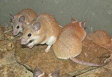
[84] In addition to these two species, subsequent studies demonstrated that Acomys cahirinus could regenerate skin and excised tissue in the ear pinna.
[87] But the regeneration therapy approach of Robert O. Becker, using electrical stimulation, has shown promising results for rats[88] and mammals in general.
[92][93] However, recent work has shown that MRL mice actually close small ear holes with scar tissue, rather than regeneration as originally claimed.
[98] Another example of physiological regeneration is the sloughing and rebuilding of a functional endometrium during each menstrual cycle in females in response to varying levels of circulating estrogen and progesterone.
Specifically, cytokine stimulation of cells leads to expression of genes that change cellular functions and suppress the immune response.
[106] Even in adult myocardium following infarction, proliferation is only found in around 1% of myocytes around the area of injury, which is not enough to restore function of cardiac muscle.