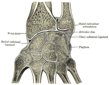
Triangular fibrocartilage
The triangular fibrocartilage disc (TFC) is an articular discus that lies on the pole of the distal ulna.
The central portion of the TFC is thin and consists of chondroid fibrocartilage; this type of tissue is often seen in structures that can bear compressive loads.
The deep components insert more anterior, into the fovea adjacent to the articular surface of the dome of the distal ulna.
[citation needed] The ligaments are composed of longitudinally oriented lamellar collagen to resist tensile loads and have a rich vascular supply to allow healing.
[citation needed] The TFCC has a substantial risk for injury and degeneration because of its anatomic complexity and multiple functions.
Application of an extension-pronation force to an axial-load wrist, such as in a fall on an outstretched hand, causes most of the traumatic injuries of the TFCC.
In cadavaric examinations, 30% to 70% of the cases had TFCC perforations and chondromalacia of the ulnar head, lunate, and triquetrum.
[1] The Palmer classification is the most recognized scheme; it divides TFCC lesions into these two categories: traumatic and degenerative.
Other symptoms patients with a TFCC injury frequently mention are: swelling, loss of grip strength, instability, and grinding or clicking sounds (crepitus) that can occur during activity of the wrist.
Note: Imaging techniques can only be relevant together with the clinical findings of a carefully performed physical examination.
The initial treatment for both traumatic and degenerative TFCC lesions, with a stable DRUJ, is conservative (nonsurgical) therapy.
[12] Closed fractures (where the skin is still intact) of the radius bone are treated non-surgically with cast; the immobilization can also help heal the TFCC.
When a tear occurs in this area of the TFC, it typically creates an unstable flap of tissue that is likely to catch on other joint surfaces.
Arthroscopic debridement as a treatment for degenerative TFC tears associated with positive ulnar variance, unfortunately, show poor results.
[citation needed] Open surgery is usually required for degenerative or more complex TFCC injuries, or if additional damage to the wrist or forearm caused instability or displacement.
[citation needed] This article incorporates text in the public domain from page 325 of the 20th edition of Gray's Anatomy (1918)