Trogocytosis
[2] The molecular reorganization occurring at the interface between the lymphocyte and the antigen-presenting cell during conjugation is also called "immunological synapse".
Like in lymphocytes, trogocytosis occurs with PMN (polymorphonuclear leukocytes, also known as granulocytes) and is associated with effective ADCC (Antibody dependent cell mediated cytotoxicity).
Outside the immune system, similar transfer of membrane fragments have been documented between sperm and oocytes, a process thought to contribute to gamete fusion.
[7] Lately the term has been attributed to macrophages, such as the CNS resident microglia, which are able to partially remove small portions of neuronal axons during postnatal development.
Such gained plasma membrane fragments could also contribute to the capacity to proliferate, because lipids are highly energetic claiming components to establish.
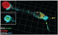
An example is rituximab, a therapeutic antibody used to treat chronic lymphocytic leukemia, recognizes the CD20 molecule expressed by tumor cells and leads to their elimination.
TRAP assays have been successfully used to detect T cell responses against viral infections,[19] cancer,[20] autoimmune diseases[21] and vaccines.