Ultrasonography of chronic venous insufficiency of the legs
Ultrasonography of suspected or previously confirmed chronic venous insufficiency of leg veins is a risk-free, non-invasive procedure.
As with heart ultrasound (echocardiography) studies, venous ultrasonography requires an understanding of hemodynamics in order to give useful examination reports.
[4][5] No preparation is normally necessary for this examination, but if a complementary study of abdominal veins is also required, the patient will be asked to fast for 12 hours beforehand.
[6][7] The ultrasound equipment must be sufficiently high-quality to give a correct image-processing result, which can then provide invaluable information, mainly at the superficial level.
Doppler measurements which trace the echoes of the generated soundwaves received by the probe, enable the direction and velocity of the blood flow to be depicted.
Also, the proper use of the scanner calls for a high level of expertise, so the examiner must be well qualified and experienced in order to give effective results.
In contrast to arterial ultrasonography, the wall of the vein is not relevant and importance is given to the hemodynamic conclusions that the examiner can obtain in order to provide a valuable report.
[10][11] Specialized training in venous ultrasonography is not undertaken in some countries, which undermines best practice, mainly when varicose veins need to be examined.
Tissue in the body will offer varying degrees of resistance, known as acoustic impedance, to the path of the ultrasound beam.
When the ultrasound beam meets air, or solid tissue such as bone, their impedance difference is so great that most of the acoustic energy is reflected, making it impossible to see any underlying structures.
The gel prevents air bubbles from forming between the probe and the patient's skin, and so helps the conduction of the sound waves from the transducer into the body.
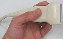
[citation needed] The probe is an ultrasonic sensor, generally known as a transducer, which functions to send and receive acoustic energy.
The reflected ultrasound is received by the probe, transformed into an electric impulse as voltage, and sent to the engine for signal processing and conversion to an image on the screen.
This low velocity is responsible for the fact that it can only be detected spontaneously with the Doppler effect on the proximal and larger femoral and iliac veins.
However, there are two exceptions: firstly, the GSV collaterals (the veins that run parallel) drain the abdominal wall and have a flow from top to bottom so that, when an examiner tests the saphenofemoral junction, a false-positive diagnosis might be made; secondly, in the flow from the sole of the foot venous network, around 10% drains to the dorsal venous arch of the foot, going therefore against the norm, from deep to superficial veins.
[22] Attention will be focused on the direction of blood flow in both venous systems, and in the perforator veins, as well as on shunt detection.
It is dependent on the examiner's expertise and training, and the interpretation of the results is subjective and reliant on an understanding of venous hemodynamics.
It travels up the leg and medial side of the thigh to reach the groin, where it drains into the common femoral vein.
[37] The small saphenous vein (SSV) runs along the posterior aspect of the leg as far as the popliteal region, in the upper calf.
For example, after a GSV stripping, laser ablation or after its ligation at the sapheno-femoral junction, the Giacomini vein will drain into the SSV, with a retrograde flow.
Where surgery, other than stripping or laser ablation is intended, the examiner will make reference to the blood flow direction in this vein, as it will be of importance.
During the muscular systole their valves close and stop any blood flow coming from the deep to the superficial veins.
When their valves become insufficient, they are responsible for a rapid deterioration in existing varicose disease and for the development of venous ulcers.
After performing this examination, the physician writes a report in which some points are crucial: [nb 4] This enables surgeons to plan interventions, in a stage known as virtual dissection.
The first ultrasound was applied to the human body for medical purposes by Dr. George Ludwig, University of Pennsylvania, in the late 1940s.
In the mid-1950s more research was undertaken by Professor Ian Donald et al., in Glasgow, which advanced the practical technology and applications of ultrasound.
In 1963, in France, Léandre Pourcelot started on his thesis, which was presented in 1964, and used pulsed Doppler for blood flow calculation as its subject.
Dr. Gene Strandness and the bioengineering group at the University of Washington, who conducted research on Doppler ultrasound as a diagnostic tool for vascular disease, published their first work in 1967.
Soon other advances in electronics and piezoelectric materials enabled further improvements, and ultrasound was quickly adopted for use in medicine due to its rapid, accurate diagnostic capabilities offering the possibility of prompt treatment.
These systems, involving digital beamforming and greater signal enhancement, have introduced new methods of interpreting and displaying data.