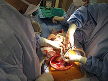
Umbilical cord
[3] The smooth muscle cells of the layer are rather poorly differentiated, contain only a few tiny myofilaments and are thereby unlikely to contribute actively to the process of post-natal closure.
Umbilical cord mesenchymal stem cells (UC-MSC) have been used clinically to treat osteoarthritis, autoimmune diseases, and multiple other conditions.
[5] The umbilical cord contains Wharton's jelly, a gelatinous substance made largely from mucopolysaccharides that protects the blood vessels inside.
The second branch (known as the ductus venosus) bypasses the liver and flows into the inferior vena cava, which carries blood towards the heart.
[10] In absence of external interventions, the umbilical cord occludes physiologically shortly after birth, explained both by a swelling and collapse of Wharton's jelly in response to a reduction in temperature and by vasoconstriction of the blood vessels by smooth muscle contraction.
[3] In contrast, the inner layer seems to serve mainly as a plastic tissue which can easily be shifted in an axial direction and then folded into the narrowing lumen to complete the closure.
[14] Within the child, the umbilical vein and ductus venosus close up, and degenerate into fibrous remnants known as the round ligament of the liver and the ligamentum venosum respectively.
Current evidence neither supports, nor refutes, delayed cutting of the cord, according to the American Congress of Obstetricians and Gynecologists (ACOG) guidelines.
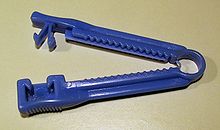
In the United States, however, where the birth occurred outside of the hospital and an emergency medical technician (EMT) clamps and cuts the cord, a longer segment up to 18 cm (7 in) in length[19][20] is left attached to the newborn.
A Cochrane review in 2013 came to the conclusion that delayed cord clamping (between one and three minutes after birth) is "likely to be beneficial as long as access to treatment for jaundice requiring phototherapy is available".
[21] In this review delayed clamping, as contrasted to early, resulted in no difference in risk of severe maternal postpartum hemorrhage or neonatal mortality, and a low Apgar score.
[21] In 2012, the American College of Obstetricians and Gynecologists officially endorsed delaying clamping of the umbilical cord for 30–60 seconds with the newborn held below the level of the placenta in all cases of preterm delivery based largely on evidence that it reduces the risk of intraventricular hemorrhage in these children by 50%.
[23] Several studies have shown benefits of delayed cord clamping: A meta-analysis[24] showed that delaying clamping of the umbilical cord in full-term neonates for a minimum of two minutes following birth is beneficial to the newborn in giving improved hematocrit, iron status as measured by ferritin concentration and stored iron, as well as a reduction in the risk of anemia (relative risk, 0.53; 95% CI, 0.40–0.70).
The entire intact umbilical cord is allowed to dry and separates on its own (typically on the 3rd day after birth), falling off and leaving a healed umbilicus.
[31] The Royal College of Obstetricians and Gynaecologists has warned about the risks of infection as the decomposing placenta tissue becomes a nest for infectious bacteria such as Staphylococcus.
[32] In one such case, a 20-hour old baby whose parents chose UCNS was brought to the hospital in an agonal state, was diagnosed with sepsis and required an antibiotic treatment for six weeks.
[25] The Royal College of Obstetricians and Gynaecologists stated in 2006 that "there is still insufficient evidence to recommend directed commercial cord blood collection and stem-cell storage in low-risk families".
[37] In the future, cord blood-derived embryonic-like stem cells (CBEs) may be banked and matched with other patients, much like blood and transplanted tissues.
In 2005, the National Academy of Sciences published an Institute of Medicine (IoM) report which recommended that expectant parents be given a balanced perspective on their options for cord blood banking.
The use of cord blood stem cells in treating conditions such as brain injury[39] and Type 1 Diabetes[40] is already being studied in humans, and earlier stage research is being conducted for treatments of stroke,[41][42] and hearing loss.
In contrast, cord blood stored in public banks is accessible to anyone with a closely matching tissue type and demonstrated need.
Engineers sometimes use the term to describe a complex or critical cable connecting a component, especially when composed of bundles of conductors of different colors, thickness and types, terminating in a single multi-contact disconnect.
Over 300 chemical toxicants have been found, including bisphenol A (BPA), tetrabromobisphenol A (TBBPA), Teflon-related perfluorooctanoic acid, galaxolide and synthetic musks among others.