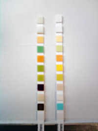
Urine test strip
The strip is then left to stand for the time necessary for the reactions to occur (usually 1 to 2 minutes), and finally the colours that appear are compared against the chromatic scale provided by the manufacturer.
An improper technique can produce false results, for example, leukocytes and erythrocytes precipitate at the bottom of the container and may not be detected if the sample is not properly mixed, and in the same way, if an excess of urine remains on the strip after it has been removed from the test sample, may cause the reagents to leak from the pads onto adjacent pads resulting in mixing and distortion of the colours.
[4] Normal reference values are not provided for urine pH as the variation is too wide and results have to be considered in the context of the other quantifiable parameters.
On the one hand it provides information regarding the balance between acid and alkali in a patient and allows identification of the substances that are present in the urine in crystalline form.
In order to differentiate pH in this wide range it is common to use a double indicator system comprising methyl red and bromothymol blue.
Reagent strip tests can detect concentrations as low as five red blood cells per microliter; however, care must be taken when comparing these figures with the actual microscopic values, because the absorbent nature of the pad attracts some of urine.
False-negative reactions can result when urine with a high specific gravity contains crenated red blood cells that do not lyse when they come in contact with the reagent pad.
[10] With the aid of routine examinations early symptoms of the following four groups can be identified: Screening parameters: Many renal and urinary tract diseases may be asymptomatic for a long period of time.
Routine urinalysis is recommended as a basic yet fundamental step in identifying renal damage and/or urinary tract disease at an early stage, especially in high-risk populations such as diabetics, the hypertensive, African Americans, Polynesians, and those with a family history.
Likewise, a technical error of allowing the reagent pad to remain in contact with the urine for a prolonged period may remove the buffer.
Highly pigmented urine and contamination of the container with quaternary ammonium compounds, detergents and antiseptics also cause false-positive readings.
The presence of blood in the urine is, of all the parameters normally tested, the one that is most closely related with traumatic damage to the kidneys or the genitourinary tract.
Under normal conditions the formation of haptoglobin-hemoglobin complexes prevents glomerular filtration, but if the hemolysis is extensive haptoglobin's uptake capacity is exceeded and hemoglobin can appear in urine.
Hemoglobinuria can be caused by hemolytic anaemia, blood transfusions, extensive burns, the bite of the recluse spider (Loxosceles), infections and strenuous exercise.
Glucose concentrations vary in an individual, and a healthy person can present with transitory glucosuria after a meal high in sugars; therefore the most representative results come from samples obtained at least two hours after food is eaten.
Elevated concentrations of ketones are not generally found in urine, as all these substances are completely metabolized, producing energy, carbon dioxide and water.
An increase in the blood concentration of ketone produces a water-electrolyte imbalance, dehydration and if not corrected, acidosis and in the end diabetic coma.
Those medicines that contain sulfhydryl groups, such as mercaptoethane sulphonate Na (Mesna) and captopril and L-DOPA can give atypical colouring.
The haemoglobin that is released after the mononuclear phagocyte system (located in the liver and spleen) withdraws old red blood cells from circulation is degraded into its components; iron, protoporphyrin and protein.
Conjugated bilirubin appears in urine when the normal degradation cycle is altered due to the obstruction of the biliary ducts or when the kidney's functional integrity is damaged.
The detection of urinary bilirubin is an early indication of liver disease and its presence or absence can be used to determine the causes of clinical jaundice.
[17] A number of substances interfere with the Ehrlich reaction on the Multistix strip: porphobilinogen, indican, p-amino salicylic acid, sulphonamide, methyldopa, procaine and chlorpromazine.
Poorly stored samples can yield false negative results as the urobilinogen suffers photo oxidation to urobilin that does not react.
Some of the gram negative bacteria species that most commonly cause urinary tract infections (Escherichia coli, Enterobacter, Klebsiella, Citrobacter and Proteus) have enzymes that reduce the nitrate present in urine to nitrite.
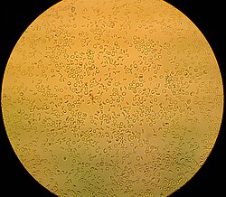
[6][20] It is normal to find up to 3 (occasionally 5) leukocytes per high power field (40X) in a urine sample, with women having slightly higher results owing to vaginal contamination.
[21] The test for leukocyte esterase is purely indicative and should not be solely relied on for diagnosis, as it does not replace microscopic or urine culture examinations.
[19] The urine test strip reaction is based on the action of leukocyte esterase in catalysing the hydrolysis of an ester of indolecarboxylic acid.
Ascorbic acid (vitamin C) is known to interfere with the oxidation reaction of the blood and glucose pad on common urine test strips.
During routine screening, if a positive test for leukocytes, blood, protein, nitrite, and a pH greater than 7 is identified, the urine sediment may be microscopically analysed to further pinpoint a diagnosis.
They can measure calcium, blood, glucose, bilirubin, urobilinogen, ketones, leukocytes, creatinine, microalbumin, pH, ascorbic acid and protein.