West Nile virus
[3] Electron microscope studies reveal a 45–50 nm virion covered with a relatively smooth protein shell; this structure is similar to the dengue fever virus, another Flavivirus.
[6][7] The lipid membrane has many roles in viral infection, including acting as signaling molecules and enhancing entry into the cell.
[4] The RNA genome is bound to capsid (C) proteins, which are 105 amino-acid residues long, to form the nucleocapsid.
The WNV genome is first translated into a polyprotein and later cleaved by virus and host proteases into separate proteins (i.e. NS1, C, E).
[24] The rest of the virus is assembled along the endoplasmic reticulum and through the Golgi apparatus, and results in non-infectious immature virions.
[29] Studies of phylogenetic lineages have determined that WNV emerged as a distinct virus around 1000 years ago.
A 2007 fatal case in a killer whale in Texas broadened the known host range of West Nile virus to include cetaceans.
[33] Since the first North American cases in 1999, the virus has been reported throughout the United States, Canada, Mexico, the Caribbean, and Central America.
[34] Both the American and Israeli strains are marked by high mortality rates in infected avian populations; the presence of dead birds—especially Corvidae—can be an early indicator of the arrival of the virus.
[36][37] Some birds, including the American crow (Corvus brachyrhynchos), blue jay (Cyanocitta cristata) and greater sage-grouse (Centrocercus urophasianus), are killed by the infection, but others survive.
[40][41] Brown thrashers (Toxostoma rufum), gray catbirds (Dumetella carolinensis), northern cardinals (Cardinalis cardinalis), northern mockingbirds (Mimus polyglottos), wood thrushes (Hylocichla mustelina) and the dove family are among the other common N. American birds in which high levels of antibodies against WNV have been found.
[38] Experimental infection has also been demonstrated with soft tick vectors, but is unlikely to be important in natural transmission.
[38][42] WNV has a broad host range, and is also known to be able to infect at least 30 mammalian species, including humans, some non-human primates,[43] horses, dogs and cats.
[35][36][40][44] Some infected humans and horses experience disease but dogs and cats rarely show symptoms.
[36] Reptiles and amphibians can also be infected, including some species of crocodiles, alligators, snakes, lizards and frogs.
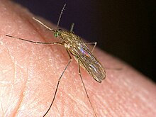
[38] In the normal rural or enzootic transmission cycle, the virus alternates between the bird reservoir and the mosquito vector.
[41][50][51] The virus can also rarely be spread through blood transfusions, organ transplants, or from mother to baby during pregnancy, delivery, or breastfeeding.
[38] According to the Centers for Disease Control and Prevention, infection with West Nile Virus is seasonal in temperate zones.
[58] All ages are equally likely to be infected but there is a higher amount of death and neuroinvasive West Nile Virus in people 60–89 years old.
When a person is in an area that has WNV, it is important to avoid outdoor activity, and if they go outside they should use a mosquito repellent with DEET.
Furthermore, higher winter temperatures and warmer spring may lead to larger summer mosquito populations, increasing the risk for WNV.
Similarly, rainfall may also drive mosquito replication rates and affect the seasonality and geographic variations of the virus.
[63] Mosquitoes have extremely wide environmental tolerances and a nearly ubiquitous geographical distribution, being present on all major land masses except Antarctica and Iceland.
Nevertheless, changes in climate and land use on ecological timescales can variously expand or fragment their distribution patterns, raising consequent concerns for human health.