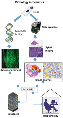
Digital pathology
[1] This field has applications in diagnostic medicine and aims to achieve more efficient and cost-effective diagnoses, prognoses, and disease predictions through advancements in machine learning and artificial intelligence in healthcare.
QuPath[9] is another such open source software, which is often used for digital pathology applications because it offers a powerful set of tools for working with whole slide images.
Image segmentation and classification algorithms, often implemented using deep neural networks, are used to identify medically significant regions and objects on digital slides.
A GPU acceleration software for pathology imaging analysis, cross-comparing spatial boundaries of a huge amount of segmented micro-anatomic objects has been developed.
[11] The core algorithm of PixelBox in this software has been adopted in Fixstars' Geometric Performance Primitives (GPP) library[12] as a part of NVIDIA Developer, which is a production geometry engine for advanced graphical information systems, electronic design automation, computer vision and motion planning solutions.
[16] The approval was based on a multi-center study of 1,992 cases in which whole-slide imaging (WSI) was shown to be non-inferior to microscopy across a wide range of surgical pathology specimens, sample types and stains.
The strongest ROI justification includes improved quality of healthcare, increased efficiency for pathologists, and reduced costs in handling glass slides.
[20] The College of American Pathologists has published a guideline with minimal requirements for validation of whole slide imaging systems for diagnostic purposes in human pathology.
Multiplexed imaging (staining multiple markers on the same slide) allows pathologists to understand finer distribution of cell-types and their relative locations.
[25] An understanding of the spatial distribution of cell-types or markers and pathways they express, can allow for prescription of targeted drugs or build combinational therapies in a personalized manner.