Abdominal ultrasonography
Abdominal ultrasound examinations are performed by gastroenterologists or other specialists in internal medicine, radiologists, or sonographers trained for this procedure.
Ultrasound can also be used if there is suspicion of enlargement of one or more organs, such as used in screening for abdominal aortic aneurysm, investigation for splenomegaly or urinary retention.
[4] In cases of infectious mononucleosis, splenomegaly is a common symptom, and health care providers may consider using abdominal ultrasonography to get insight into a person's condition.
Tumor characterization using the ultrasound method will be based on the following elements: consistency (solid, liquid, mixed), echogenicity, structure appearance (homogeneous or heterogeneous), delineation from adjacent liver parenchyma (capsular, imprecise), elasticity, posterior acoustic enhancement effect, the relation with neighboring organs or structures (displacement, invasion), vasculature (presence and characteristics on Doppler ultrasonography and contrast-enhanced ultrasound (CEUS).
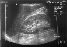
[citation needed] Ultrasonography of the kidneys is essential in the diagnosis and management of kidney-related diseases.
[7] Advantages of ultrasound imaging of abdominal structures are that the procedure can be performed quickly, bed-side, involves no exposure to X-rays (which makes it useful in pregnant patients, for example) and is inexpensive compared to other often-used techniques such as computed tomography (CT scan) of the abdomen.
[citation needed] The imaging occurs real-time and without sedation, so that the influence of movements can be assessed quickly.
Through the abdominal wall, organs inside the pelvis can be seen, such as the urinary bladder or the ovaries and uterus in women.
The liver can be imaged by swiping the probe sagittally from medial to lateral at the subcoastal region.
Common Bile Duct: Nondilated measuring 1.3 mm at the level of the porta hepatis.