Adeno-associated virus
[1][2] Several features make AAV an attractive candidate for creating viral vectors for gene therapy, and for the creation of isogenic human disease models.
[5] In March 2023, a series of Nature papers detected high titres of adeno-associated virus 2 (AAV2), alongside adenovirus and herpesvirus, in samples from a wave of childhood hepatitis.
[6] One paper suggested that AAV2 co-infection may contribute to more serious liver disease than infection with only adeno- or herpesviruses and that the causal link remains to be established.
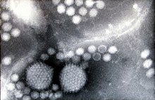
[7] The adeno-associated virus (AAV), previously thought to be a contaminant in adenovirus preparations, was first identified as a dependoparvovirus in the 1960s in the laboratories of Bob Atchison at Pittsburgh and Wallace Rowe at NIH.
[11] AAVs also present very low immunogenicity, seemingly restricted to generation of neutralizing antibodies, while they induce no clearly defined cytotoxic response.
The cloning capacity of the vector is relatively limited and most therapeutic genes require the complete replacement of the virus's 4.8 kilobase genome.
The genome comprises ITRs at both ends of the DNA strand, and two open reading frames (ORFs): rep and cap.
[37] The feature of these sequences that gives them this property is their ability to form a hairpin, which contributes to so-called self-priming that allows primase-independent synthesis of the second DNA strand.
With this assumption many methods were established for efficient production of recombinant AAV (rAAV) vectors containing a reporter or therapeutic gene.
[41][42][43][44] On the "left side" of the genome there are two promoters called p5 and p19, from which two overlapping messenger ribonucleic acids (mRNAs) of different length can be produced.
[50] The AAV capsid is composed of a mixture of VP1, VP2, and VP3 totaling 60 monomers arranged in icosahedral symmetry in a ratio of 1:1:10,[51] with an empty mass of approximately 3.8 MDa.
However, upstream of that codon in the same open reading frame lies an ACG sequence (encoding threonine) which is surrounded by an optimal Kozak context.
[59] The unique fragment at the N terminus of VP1 protein was shown to possess the phospholipase A2 (PLA2) activity, which is probably required for the releasing of AAV particles from late endosomes.
[57] More recently, however, Warrington et al. showed VP2 to be unnecessary for the complete virus particle formation and an efficient infectivity, and also presented that VP2 can tolerate large insertions in its N terminus, while VP1 can not, probably because of the PLA2 domain presence.
[68] AAV capsid proteins contain 12 hypervariable surface regions, with most variability occurring in the threefold proximal peaks, but the parvovirus genome in general presents highly conserved replication and structural genes across serotypes.
Tissue specificity is determined by the capsid serotype and pseudotyping of AAV vectors to alter their tropism range will likely be important to their use in therapy.
[78][79][80] These study results have been disputed by Qiu, Handa, et al.[81] HSPG functions as the primary receptor, though its abundance in the extracellular matrix can scavenge AAV particles and impair the infection efficiency.
For example, AAV4 and AAV5 transduction can be inhibited by soluble sialic acids (of different form for each of these serotypes),[92] and AAV5 was shown to enter cells via the platelet-derived growth factor receptor.
Such modifications include new tropisms to target specific tissues, and modified surface residues to evade detection by the immune system.
[96] AAV is of particular interest to gene therapists due to its apparent limited capacity to induce immune responses in humans, a factor which should positively influence vector transduction efficiency while reducing the risk of any immune-associated pathology.
Intravenous administration in mice causes transient production of pro-inflammatory cytokines and some infiltration of neutrophils and other leukocytes into the liver, which seems to sequester a large percentage of the injected viral particles.
[101] In-vivo studies indicate that AAV vectors interact with the Toll-like receptor (TLR)9- and TLR2-MyD88 pathways to trigger the innate immune response by stimulating the production of interferons.
[105] The virus is known to instigate robust humoral immunity in animal models and in the human population, where up to 80% of individuals are thought to be seropositive for AAV2.
As well as persistent AAV specific antibody levels, it appears from both prime-boost studies in animals and from clinical trials that the B-cell memory is also strong.
[106] Clinical trials using an AAV2-based vector to treat haemophilia B seem to indicate that targeted destruction of transduced cells may be occurring.
It was then shown that AAV replication can be facilitated by selected proteins derived from the adenovirus genome,[111][112] by other viruses such as HSV[113] or vaccinia, or by genotoxic agents, such as UV irradiation or hydroxyurea.
[114][115][116] Depending on the presence or absence of a helper virus, the life cycle of AAV follows either a lytic or lysogenic pathway, respectively.
In the absence of helper virus or genotoxic factors, AAV DNA can either integrate into the host genome or persist in episomal form.
In mice, the AAV genome has been observed persisting for long periods of time in quiescent tissues, such as skeletal muscles, in episomal form (a circular head-to-tail conformation).