Spinal cord
The cervical enlargement, stretching from the C4 to T1 vertebrae, is where sensory input comes from and motor output goes to the arms and trunk.
The spinal cord is continuous with the caudal portion of the medulla, running from the base of the skull to the body of the first lumbar vertebra.
The dorsal roots are afferent fascicles, receiving sensory information from the skin, muscles, and visceral organs to be relayed to the brain.
[5] The spinal cord (and brain) are protected by three layers of tissue or membranes called meninges, that surround the canal.
In cross-section, the peripheral region of the cord contains neuronal white matter tracts containing sensory and motor axons.
The cauda equina ("horse's tail") is a collection of nerves inferior to the conus medullaris that continue to travel through the vertebral column to the coccyx.
The cauda equina forms because the spinal cord stops growing in length at about age four, even though the vertebral column continues to lengthen until adulthood.
The inferior part of the vertebral canal is filled with cerebrospinal fluid and the space is called the lumbar cistern.
[8] Within the central nervous system (CNS), nerve cell bodies are generally organized into functional clusters, called nuclei, their axons are grouped into tracts.
They form anastomoses (connections) via the anterior and posterior segmental medullary arteries, which enter the spinal cord at various points along its length.
[11] The actual blood flow caudally through these arteries, derived from the posterior cerebral circulation, is inadequate to maintain the spinal cord beyond the cervical segments.
[11] These intercostal and lumbar radicular arteries arise from the aorta, provide major anastomoses and supplement the blood flow to the spinal cord.
During the maturation of the neural tube, its lateral walls thicken and form a longitudinal groove called the sulcus limitans.
Opposing gradients of such morphogens as BMP and SHH form different domains of dividing cells along the dorsal ventral axis.
As the dorsal and ventral column cells proliferate, the lumen of the neural tube narrows to form the small central canal of the spinal cord.
The netrins act as chemoattractants to decussation of pain and temperature sensory neurons in the alar plate across the anterior white commissure, where they then ascend towards the thalamus.
Secondary axons from the medial lemniscus finally terminate in the ventral posterolateral nucleus (VPLN) of the thalamus, where they synapse with tertiary neurons.
Its primary neurons axons enter the spinal cord and then ascend one to two levels before synapsing in the substantia gelatinosa.
After synapsing, secondary axons decussate and ascend in the anterior lateral portion of the spinal cord as the spinothalamic tract.
From the levels of L2 to T1, proprioceptive information enters the spinal cord and ascends ipsilaterally, where it synapses in Clarke's nucleus.
The secondary neuronal axons continue to ascend ipsilaterally and then pass into the cerebellum via the inferior cerebellar peduncle.
From above T1, proprioceptive primary axons enter the spinal cord and ascend ipsilaterally until reaching the accessory cuneate nucleus, where they synapse.
[2] In mice, a projection exists from primary and secondary somatosensory cortex to interneurons in laminae III-V of the lumbar spinal cord that aids the detection of light touch.
[20] In humans, research finds prior knowledge about when sensory input will happens top-down modulates spinal activity and does so with responses as early at 13 to 16 ms after stimulation.
[21] A congenital disorder is diastematomyelia in which part of the spinal cord is split usually at the level of the upper lumbar vertebrae.
Damage to upper motor neuron axons in the spinal cord results in a characteristic pattern of ipsilateral deficits.
Spinal stenoses at the lumbar region are usually due to disc herniation, hypertrophy of the facet joint and ligamentum flavum, osteophyte, and spondylolisthesis.
The epidural fat can be seen as low density on CT scan and high intensity on T2-weighted fast spin echo MRI images.
The spinal cord ends at the level of vertebrae L1–L2, while the subarachnoid space – the compartment that contains cerebrospinal fluid – extends down to the lower border of S2.
[22] Lumbar punctures in adults are usually performed between L3–L5 (cauda equina level) in order to avoid damage to the spinal cord.
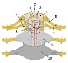
| 1 | central canal |
| 2 | posterior median sulcus |
| 3 | gray matter |
| 4 | white matter |
| 5 |
dorsal root (left),
dorsal root ganglion (right) |
| 6 | ventral root |
| 7 | fascicles |
| 8 | anterior spinal artery |
| 9 | arachnoid mater |
| 10 | dura mater |


