Bone marrow examination
Bone marrow examination is used in the diagnosis of a number of conditions, including leukemia, multiple myeloma, lymphoma, anemia, and pancytopenia.
The aspirate yields semi-liquid bone marrow, which can be examined by a pathologist under a light microscope and analyzed by flow cytometry, chromosome analysis, or polymerase chain reaction (PCR).
Frequently, a trephine biopsy is also obtained, which yields a narrow, cylindrically shaped solid piece of bone marrow, 2 mm wide and 2 cm long (80 μL), which is examined microscopically (sometimes with the aid of immunohistochemistry) for cellularity and infiltrative processes.
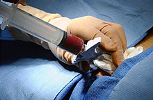
[citation needed] Bone marrow aspiration and trephine biopsy are usually performed on the back of the hipbone, or posterior iliac crest.
Then, with a twisting motion of clinician's hand and wrist, the needle is advanced through the bony cortex (the hard outer layer of the bone) and into the marrow cavity.
It is important to note that thrombocytopenia or bleeding disorders are not contraindications as long as the procedure is performed by a skilled clinician.
[3] Bone marrow aspiration and biopsy can be safely performed even in the setting of extreme thrombocytopenia (low platelet count).
[citation needed] While mild soreness lasting 12–24 hours is common after a bone marrow examination, serious complications are extremely rare.
[4] The same author collected data on over 19,000 bone marrow examinations performed in the United Kingdom in 2003, and found 16 adverse events (0.08% of total procedures), the most common of which was bleeding.