Lymphocyte
B cells respond to pathogens by producing large quantities of antibodies which then neutralize foreign objects like bacteria and viruses.
[10][11] However, the authors of original article pointed to the fact that the two studies have detected X cells by imaging microscopy and FACS as described.
[12] Additional studies are required to determine the nature and properties of X cells (also called dual expressers).
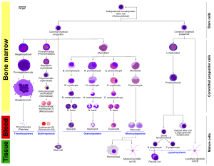
T cells migrate to the blood stream and mature in a distinct primary organ, called the thymus.
Following maturation, the lymphocytes enter the circulation and peripheral lymphoid organs (e.g. the spleen and lymph nodes) where they survey for invading pathogens and/or tumor cells.
Memory T cells remain in the peripheral tissues and circulation for an extended time ready to respond to the same antigen upon future exposure; they live weeks to several years, which is very long compared to other leukocytes.
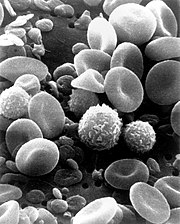
[citation needed] Microscopically, in a Wright's stained peripheral blood smear, a normal lymphocyte has a large, dark-staining nucleus with little to no eosinophilic cytoplasm.
In normal situations, the coarse, dense nucleus of a lymphocyte is approximately the size of a red blood cell (about 7 μm in diameter).
The ribosomes are involved in protein synthesis, allowing the generation of large quantities of cytokines and immunoglobulins by these cells.
Lymphoproliferative disorders (LPD) encompass a diverse group of diseases marked by uncontrolled lymphocyte production, leading to issues like lymphocytosis, lymphadenopathy, and bone marrow infiltration.
These disorders are common in immunocompromised individuals and involve abnormal proliferation of T and B cells, often resulting in immunodeficiency and immune system dysfunction.
LPDs encompass a wide array of disorders involving B-cell (e.g., chronic lymphocytic leukemia) and T-cell (e.g., Sezary syndrome) abnormalities, each presenting distinct challenges in diagnosis and management.
[22] Without the key defense that these T cells provide, the body becomes susceptible to opportunistic infections that otherwise would not affect healthy people.