Brodmann area

A Brodmann area is a region of the cerebral cortex, in the human or other primate brain, defined by its cytoarchitecture, or histological structure and organization of cells.

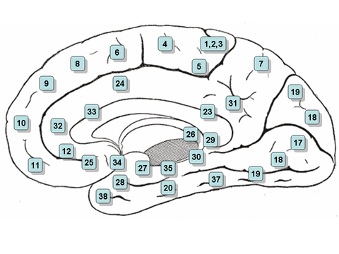
Brodmann mapped the human brain based on the varied cellular structure across the cortex and identified 52 distinct regions, which he numbered 1 to 52.

Brodmann published his maps of cortical areas in humans, monkeys, and other species in 1909,[2] along with many other findings and observations regarding the general cell types and laminar organization of the mammalian cortex.

[4] Brodmann areas have been discussed, debated, refined, and renamed exhaustively for nearly a century and remain the most widely known and frequently cited cytoarchitectural organization of the human cortex.

A simple example of this type of correspondence is the primary motor cortex, a strip of tissue running along the anterior edge of the central sulcus.

This "somatotopic" representation is not evenly distributed, however; the head, for example, is represented by a region about three times as large as the zone for the entire back and trunk.

The maps for visual areas are retinotopic, meaning that they reflect the topography of the retina: the layer of light-activated neurons lining the back of the eye.

[7] When von Bonin and Bailey constructed a brain map for the macaque monkey, they found the description of Brodmann inadequate and wrote: "Brodmann (1907), it is true, prepared a map of the human brain which has been widely reproduced, but, unfortunately, the data on which it was based was never published"[8] They instead used the cytoarchitectonic scheme of Constantin von Economo and Georg N. Koskinas published in 1925[4] which had the "only acceptable detailed description of the human cortex".

Brodmann's classification of areas of the cortex
Topography of the primary motor cortex, showing which zone controls each body part
Image mapped Brodmann Areas. Clicking on an area in the picture causes the browser to load the appropriate article. Areas 3, 1 & 2 – Primary Somatosensory Cortex Area 4 – Primary Motor Cortex Area 5 – Somatosensory Association Cortex Area 6 – Premotor cortex and Supplementary Motor Cortex (Secondary Motor Cortex)(Supplementary motor area Area 7 – Somatosensory Association Cortex Area 8 – Includes Frontal eye fields Area 9 – Dorsolateral prefrontal cortex Area 10 – Anterior prefrontal cortex Area 11 – Orbitofrontal area Area 17 – Primary visual cortex (V1) Area 18 – Secondary visual cortex (V2) Area 19 – Associative visual cortex (V3) Area 20 – Inferior temporal gyrus Area 21 – Middle temporal gyrus Area 22 – Superior temporal gyrus Area 37 – Fusiform gyrus Area 38 – PULA Area 39 – Angular gyrus Area 40 – Supramarginal gyrus Area 41 – Primary and Auditory Association Cortex Area 42 – Primary and Auditory Association Cortex Area 43 – Primary gustatory cortex Area 44 – pars opercularis, part of Broca's area Area 45 – pars triangularis, Broca's area Area 46 – Dorsolateral prefrontal cortex Area 47 – Inferior prefrontal gyrus Image Map
Image mapped Brodmann Areas . Clicking on an area in the picture causes the browser to load the appropriate article.
Image mapped Brodmann Areas. Clicking on an area in the picture causes the browser to load the appropriate article. Areas 3, 1 & 2 – Primary Somatosensory Cortex area 4 – primary motor cortex Area 5 – Somatosensory Association Cortex Area 6 – Premotor cortex and Supplementary Motor Cortex (Secondary Motor Cortex)(Supplementary motor area Area 7 – Somatosensory Association Cortex Area 8 – Includes Frontal eye fields Area 9 – Dorsolateral prefrontal cortex Area 10 – Anterior prefrontal cortex Area 11 – Orbitofrontal area Area 12 – Orbitofrontal area Area 17 – Primary visual cortex (V1) Area 18 – Secondary visual cortex (V2) Area 19 – Associative visual cortex (V3) Area 19 – Associative visual cortex (V3) Area 18 – Secondary visual cortex (V2) Area 23 – Ventral Posterior cingulate cortex Area 24 – Ventral Anterior cingulate cortex Area 25 – Subgenual cortex (part of the Ventromedial prefrontal cortex) Area 26 – Ectosplenial portion of the retrosplenial region of the cerebral cortex Area 27 – Piriform cortex Area 28 – Posterior Entorhinal Cortex Area 29 – Retrosplenial cingulate cortex Area 30 – Part of cingulate cortex Area 31 – Dorsal Posterior cingulate cortex Area 32 – Dorsal anterior cingulate cortex Area 33 – Part of anterior cingulate cortex Area 34 – Anterior Entorhinal Cortex (on the Parahippocampal gyrus) Area 35 – Perirhinal cortex (on the Parahippocampal gyrus) Area 20 – Inferior temporal gyrus Area 37 – Fusiform gyrus Area 38 – Temporopolar area Image Map
Image mapped Brodmann Areas . Clicking on an area in the picture causes the browser to load the appropriate article.